Joint Development - Temporomandibular Joint: Difference between revisions
mNo edit summary |
mNo edit summary |
||
| Line 1: | Line 1: | ||
{{Header}} | {{Header}} | ||
==Introduction== | ==Introduction== | ||
The | The {{temporomandibular joint}} (TMJ) is a bilateral synovial articulation between the ends of the mandible (lower jaw) and temporal bone ( part of the {{skull}}). Both mastication and facial muscles are attached to the lower jaw. | ||
The earliest review of human embryonic and fetal TMJ development is by Symons in 1952.{{#pmid:12980883|PMID12980883}} | The earliest review of human embryonic and fetal TMJ development is by Symons in 1952.{{#pmid:12980883|PMID12980883}} | ||
Revision as of 07:45, 9 May 2018
| Embryology - 23 Apr 2024 |
|---|
| Google Translate - select your language from the list shown below (this will open a new external page) |
|
العربية | català | 中文 | 中國傳統的 | français | Deutsche | עִברִית | हिंदी | bahasa Indonesia | italiano | 日本語 | 한국어 | မြန်မာ | Pilipino | Polskie | português | ਪੰਜਾਬੀ ਦੇ | Română | русский | Español | Swahili | Svensk | ไทย | Türkçe | اردو | ייִדיש | Tiếng Việt These external translations are automated and may not be accurate. (More? About Translations) |
Introduction
The temporomandibular joint (TMJ) is a bilateral synovial articulation between the ends of the mandible (lower jaw) and temporal bone ( part of the skull). Both mastication and facial muscles are attached to the lower jaw.
The earliest review of human embryonic and fetal TMJ development is by Symons in 1952.[1]
In the adult, the region where two skeletal bones meet and articulate is called a "joint", that are classified based upon their: anatomical structure, mobility and shape. In the embryo, the majority of the vertebrate skeleton is initially formed as a cartilage template, that is later replaced by bone except at the interface between two adjacent bones, leaving in the adult a layer of cartilage in this region. The musculoskeletal system consists of skeletal muscle, bone, and cartilage and is mainly mesoderm in origin with some neural crest contribution.
| Joint Links: joint | synovial joint | temporomandibular joint | musculoskeletal | cartilage | Category:Joint | ||
|
Historic Embryology:
- Symons NB. The development of the human mandibular joint. (1952) J Anat. 86(3):326-32. PMID 12980883
- Whillis J. The development of synovial joints. (1940) J Anat. 74(Pt 2): 277-283. PMID: 17104813
Some Recent Findings
|
| More recent papers |
|---|
 This table allows an automated computer search of the external PubMed database using the listed "Search term" text link.
More? References | Discussion Page | Journal Searches | 2019 References | 2020 References Search term: Temporomandibular Joint Development <pubmed limit=5>Temporomandibular Joint Development</pubmed> |
Fetal Development
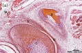
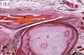
These images are from a recent article on fetal human TMJ development.[3] Weeks are GA.

Week 10 Human fetus (55 mm GL) |

Week 12 Human fetus (95 mm GL) |

Week 14 Human fetus (125 mm GL) |

Week 18 Human fetus (175 mm GL). |

Week 28 Human fetus (233 mm GL). |

Week 32 Human fetus (300 mm GL) |
Joint Types
Classification
- Fibrous (synarthrodial) - immoveable joints found in cranial vault and teeth
- Cartilagenous (synchondroses and sympheses) - partially moveable joints
- Synovial (diarthrosis) - freely moveable joints are the most common found in the skeleton
Movement

- Hinge - (elbow and knee) Flexion/Extension
- Pivot - (neck, atlas and axis bones) Rotation of one bone around another
- Ball and Socket - (shoulder and hip)
- Saddle - (thumb)
- Condyloid - (wrist joints)
- Gliding - (intercarpal joints) Gliding movements
Molecular
Indian Hedgehog (IHH)
- Human cytogenetic location - 2q35
- 336 amino acid protein.
- Sonic Hedgehog (SHH) and IHH N-terminals share 91.4% identity, C-terminal halves significantly different.
- expressed in the prehypertrophic chondrocytes of cartilage elements.
- Links: Sonic hedgehog | OMIM IHH
Short Stature Homeobox 2 (SHOX2)
- Human cytogenetic location - 3q25.32
Links: OMIM SHOX2
Temporomandibular Abnormalities
References
- ↑ SYMONS NB. (1952). The development of the human mandibular joint. J. Anat. , 86, 326-32. PMID: 12980883
- ↑ Li X, Liang W, Ye H, Weng X, Liu F, Lin P & Liu X. (2015). Overexpression of Indian hedgehog partially rescues short stature homeobox 2-overexpression-associated congenital dysplasia of the temporomandibular joint in mice. Mol Med Rep , 12, 4157-64. PMID: 26096903 DOI.
- ↑ 3.0 3.1 Alvez CS, Carvalho de Moraes LO, Marques SR, Tedesco RC, Harb LJ, Rodríguez-Vázquez JF, Mérida-Velasco JR & Alonso LG. (2014). Analysis by Light, Scanning, and Transmission Microscopy of the Intima Synovial of the Temporomandibular Joint of Human Fetuses during the Development. Anat Res Int , 2014, 732720. PMID: 24527214 DOI.
Online Textbooks
Developmental Biology Gilbert, Scott F. Sunderland (MA): Sinauer Associates, Inc. ; c2000 Forming the joints
Reviews
Kaneyama K, Segami N & Hatta T. (2008). Congenital deformities and developmental abnormalities of the mandibular condyle in the temporomandibular joint. Congenit Anom (Kyoto) , 48, 118-25. PMID: 18778456 DOI.
Abramowicz S, Marshall CJ, Dolwick MF & Cohen D. (2007). Vascular malformation of the temporomandibular joint: report of a case and review of the literature. Oral Surg Oral Med Oral Pathol Oral Radiol Endod , 103, 203-6. PMID: 17234536 DOI.
SYMONS NB. (1952). The development of the human mandibular joint. J. Anat. , 86, 326-32. PMID: 12980883
Articles
Mérida-Velasco JR, Rodríguez-Vázquez JF, Mérida-Velasco JA, Sánchez-Montesinos I, Espín-Ferra J & Jiménez-Collado J. (1999). Development of the human temporomandibular joint. Anat. Rec. , 255, 20-33. PMID: 10321990
Perry HT, Xu Y & Forbes DP. (1985). The embryology of the temporomandibular joint. Cranio , 3, 125-32. PMID: 3855934
Search PubMed
Search Pubmed: Temporomandibular Joint Development
Additional Images

Adult axial skeleton

Bone structure

Endochondral bone

Fetal head lateral (12 weeks)

Fetal head medial (12 weeks)

Fetal head section (12 weeks)
Fetal temporomandibular joint 10 weeks
Fetal temporomandibular joint 12 weeks


External Links
External Links Notice - The dynamic nature of the internet may mean that some of these listed links may no longer function. If the link no longer works search the web with the link text or name. Links to any external commercial sites are provided for information purposes only and should never be considered an endorsement. UNSW Embryology is provided as an educational resource with no clinical information or commercial affiliation.
Glossary Links
- Glossary: A | B | C | D | E | F | G | H | I | J | K | L | M | N | O | P | Q | R | S | T | U | V | W | X | Y | Z | Numbers | Symbols | Term Link
Cite this page: Hill, M.A. (2024, April 23) Embryology Joint Development - Temporomandibular Joint. Retrieved from https://embryology.med.unsw.edu.au/embryology/index.php/Joint_Development_-_Temporomandibular_Joint
- © Dr Mark Hill 2024, UNSW Embryology ISBN: 978 0 7334 2609 4 - UNSW CRICOS Provider Code No. 00098G