Joint Development - Temporomandibular Joint: Difference between revisions
mNo edit summary |
mNo edit summary |
||
| Line 15: | Line 15: | ||
|-bgcolor="F5FAFF" | |-bgcolor="F5FAFF" | ||
| | | | ||
* '''Overexpression of Indian hedgehog partially rescues short stature homeobox 2-overexpression‑associated congenital dysplasia of the temporomandibular joint in mice'''<ref name="PMID26096903"><pubmed>26096903</pubmed></ref> "The role of short stature homeobox 2 (shox2) in the development and homeostasis of the temporomandibular joint (TMJ) has been well documented. Shox2 is known to be expressed in the progenitor cells and perichondrium of the developing condyle. A previous study by our group reported that overexpression of shox2 leads to congenital dysplasia of the TMJ via downregulation of the Indian hedgehog (Ihh) signaling pathway, which is essential for embryonic disc primordium formation and mandibular condylar growth. To determine whether overexpression of Ihh may rescue the overexpression of shox2 leading to congenital dysplasia of the TMJ, a mouse model in which Ihh and shox2 were overexpressed (Wnt1-Cre; pMes-stop shox2; pMes-stop Ihh mice) was utilized to assess the consequences of this overexpression on TMJ development during post-natal life. ... These combinatory cellular and molecular defects appeared to account for the observed congenital dysplasia of TMJ, suggesting that overexpression of Ihh partially rescued shox2 overexpression‑associated congenital dysplasia of the TMJ in mice." [[Mouse Development]] | * '''Overexpression of Indian hedgehog partially rescues short stature homeobox 2-overexpression‑associated congenital dysplasia of the temporomandibular joint in mice'''<ref name="PMID26096903"><pubmed>26096903</pubmed>| [http://www.spandidos-publications.com/10.3892/mmr.2015.3959 Mol Med Rep.]</ref> "The role of short stature homeobox 2 (shox2) in the development and homeostasis of the temporomandibular joint (TMJ) has been well documented. Shox2 is known to be expressed in the progenitor cells and perichondrium of the developing condyle. A previous study by our group reported that overexpression of shox2 leads to congenital dysplasia of the TMJ via downregulation of the Indian hedgehog (Ihh) signaling pathway, which is essential for embryonic disc primordium formation and mandibular condylar growth. To determine whether overexpression of Ihh may rescue the overexpression of shox2 leading to congenital dysplasia of the TMJ, a mouse model in which Ihh and shox2 were overexpressed (Wnt1-Cre; pMes-stop shox2; pMes-stop Ihh mice) was utilized to assess the consequences of this overexpression on TMJ development during post-natal life. ... These combinatory cellular and molecular defects appeared to account for the observed congenital dysplasia of TMJ, suggesting that overexpression of Ihh partially rescued shox2 overexpression‑associated congenital dysplasia of the TMJ in mice." [[Mouse Development]] | ||
* '''Analysis by Light, Scanning, and Transmission Microscopy of the Intima Synovial of the Temporomandibular Joint of Human Fetuses during the Development'''<ref name="PMID24527214"><pubmed>24527214</pubmed>| [http://www.hindawi.com/journals/ari/2014/732720 Anat Res Int.]</ref> "To characterize morphologically and ultrastructurally using light microscopy, the scanning electron microscopy and transmission electron microscopy the intima synovial of the temporomandibular joint (TMJ) of human fetuses between the 10th and the 38th week of development. Materials and Methods. The TMJ was dissected bilaterally in 37 human fetuses belonging to the Institute of Embryology of the University Complutense of Madrid and of the Federal University of São Paulo. Results. The outcome by light microscopy showed the morphology of the TMJ and that the formation of inferior joint cavity precedes the superior joint cavity and the presence of blood vessels in the synovial. Conclusion. By scanning and transmission electron microscopy we observed the presence of two well-defined cell types in the intima layer of synovial of the TMJ of human fetuses, macrophage-like type A cell and fibroblast-like type B cell, and the presence of the a third cell type, defined by the name of intermediate lining cell in the intima layer of the synovial." | * '''Analysis by Light, Scanning, and Transmission Microscopy of the Intima Synovial of the Temporomandibular Joint of Human Fetuses during the Development'''<ref name="PMID24527214"><pubmed>24527214</pubmed>| [http://www.hindawi.com/journals/ari/2014/732720 Anat Res Int.]</ref> "To characterize morphologically and ultrastructurally using light microscopy, the scanning electron microscopy and transmission electron microscopy the intima synovial of the temporomandibular joint (TMJ) of human fetuses between the 10th and the 38th week of development. Materials and Methods. The TMJ was dissected bilaterally in 37 human fetuses belonging to the Institute of Embryology of the University Complutense of Madrid and of the Federal University of São Paulo. Results. The outcome by light microscopy showed the morphology of the TMJ and that the formation of inferior joint cavity precedes the superior joint cavity and the presence of blood vessels in the synovial. Conclusion. By scanning and transmission electron microscopy we observed the presence of two well-defined cell types in the intima layer of synovial of the TMJ of human fetuses, macrophage-like type A cell and fibroblast-like type B cell, and the presence of the a third cell type, defined by the name of intermediate lining cell in the intima layer of the synovial." | ||
|} | |} | ||
Revision as of 11:53, 3 July 2015
| Embryology - 20 Apr 2024 |
|---|
| Google Translate - select your language from the list shown below (this will open a new external page) |
|
العربية | català | 中文 | 中國傳統的 | français | Deutsche | עִברִית | हिंदी | bahasa Indonesia | italiano | 日本語 | 한국어 | မြန်မာ | Pilipino | Polskie | português | ਪੰਜਾਬੀ ਦੇ | Română | русский | Español | Swahili | Svensk | ไทย | Türkçe | اردو | ייִדיש | Tiếng Việt These external translations are automated and may not be accurate. (More? About Translations) |
Introduction
The Temporomandibular Joint (TMJ) is a bilateral synovial articulation between the ends of the mandible (lower jaw) and temporal bone ( part of the skull).
In the adult, the region where two skeletal bones meet and articulate is called a "joint", that are classified based upon their: anatomical structure, mobility and shape.
In the embryo, the majority of the vertebrate skeleton is initially formed as a cartilage template, that is later replaced by bone except at the interface between two adjacent bones, leaving in the adult a layer of cartilage in this region. The musculoskeletal system consists of skeletal muscle, bone, and cartilage and is mainly mesoderm in origin with some neural crest contribution.
| Joint Links: joint | synovial joint | temporomandibular joint | musculoskeletal | cartilage | Category:Joint | ||
|
Some Recent Findings
|
| More recent papers |
|---|
 This table allows an automated computer search of the external PubMed database using the listed "Search term" text link.
More? References | Discussion Page | Journal Searches | 2019 References | 2020 References Search term: Temporomandibular Joint Development <pubmed limit=5>Temporomandibular Joint Development</pubmed> |
Fetal Development
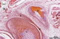
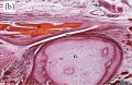
These images are from a recent article on fetal human TMJ development.[2] Weeks are GA.

Week 10 Human fetus (55 mm GL) |

Week 12 Human fetus (95 mm GL) |

Week 14 Human fetus (125 mm GL) |

Week 18 Human fetus (175 mm GL). |

Week 28 Human fetus (233 mm GL). |

Week 32 Human fetus (300 mm GL) |
Joint Types
Classification
- Fibrous (synarthrodial) - immoveable joints found in cranial vault and teeth
- Cartilagenous (synchondroses and sympheses) - partially moveable joints
- Synovial (diarthrosis) - freely moveable joints are the most common found in the skeleton
Movement

- Hinge - (elbow and knee) Flexion/Extension
- Pivot - (neck, atlas and axis bones) Rotation of one bone around another
- Ball and Socket - (shoulder and hip)
- Saddle - (thumb)
- Condyloid - (wrist joints)
- Gliding - (intercarpal joints) Gliding movements
Temporomandibular Abnormalities
References
- ↑ <pubmed>26096903</pubmed>| Mol Med Rep.
- ↑ 2.0 2.1 <pubmed>24527214</pubmed>| Anat Res Int.
Online Textbooks
Developmental Biology Gilbert, Scott F. Sunderland (MA): Sinauer Associates, Inc. ; c2000 Forming the joints
Reviews
<pubmed></pubmed>
Articles
<pubmed></pubmed>
Search PubMed
S Search Pubmed: Joint Development
Additional Images

Adult axial skeleton

Bone structure

Endochondral bone

Fetal head lateral (12 weeks)

Fetal head medial (12 weeks)

Fetal head section (12 weeks)
Fetal temporomandibular joint 10 weeks
Fetal temporomandibular joint 12 weeks


External Links
External Links Notice - The dynamic nature of the internet may mean that some of these listed links may no longer function. If the link no longer works search the web with the link text or name. Links to any external commercial sites are provided for information purposes only and should never be considered an endorsement. UNSW Embryology is provided as an educational resource with no clinical information or commercial affiliation.
Glossary Links
- Glossary: A | B | C | D | E | F | G | H | I | J | K | L | M | N | O | P | Q | R | S | T | U | V | W | X | Y | Z | Numbers | Symbols | Term Link
Cite this page: Hill, M.A. (2024, April 20) Embryology Joint Development - Temporomandibular Joint. Retrieved from https://embryology.med.unsw.edu.au/embryology/index.php/Joint_Development_-_Temporomandibular_Joint
- © Dr Mark Hill 2024, UNSW Embryology ISBN: 978 0 7334 2609 4 - UNSW CRICOS Provider Code No. 00098G