Intermediate - Heart Valves
| Begin Intermediate: | Primordial Heart Tube | Heart Tube Looping | Atrial Ventricular Septation | Outflow Tract | Heart Valves | Cardiac Abnormalities | Vascular Overview |
| Cardiac Embryology | Begin Basic | Begin Intermediate | Begin Advanced |
There are four valves in the adult heart, depicted below. There are two AV valves which comprise leaflets as well as the structures that tether these leaflets to the ventricular walls. The aortic and pulmonary valves, termed the semilunar valves, are located in the aorta and pulmonary trunk respectively. They are each made of three cusps.
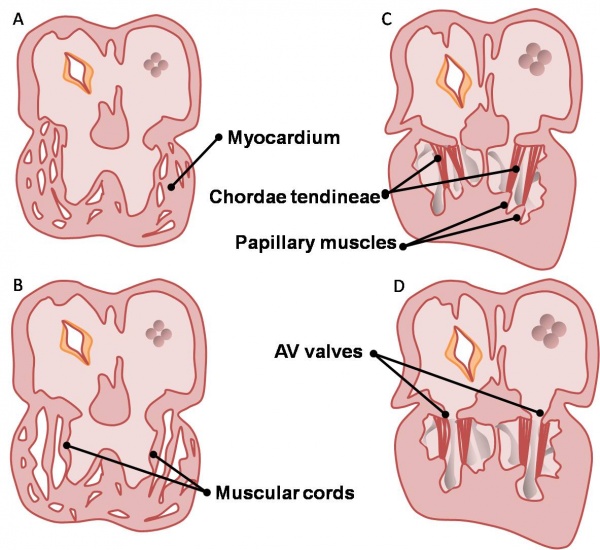
The AV valves begin to form between the fifth and eighth weeks of development. The left AV valve has anterior and posterior leaflets and is termed the bicuspid or mitral valve. The right AV valve has a third, small, septal cusp and thus is called the tricuspid valve. The valve leaflets are attached to the ventricular walls by thin fibrous chords: the chordae tendineae, which insert into small muscles attached to the ventricle wall: the papillary muscles. These structures are sculpted from the ventricular wall (see left).
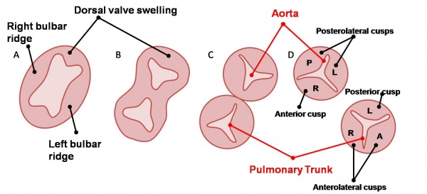
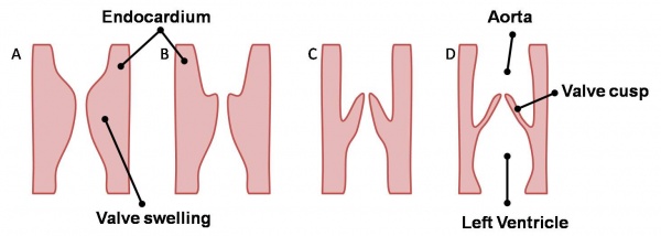
The semilunar valves are formed from the bulbar ridges and subendocardial valve tissue. The primordial semilunar valve consists of a mesenchymal core covered by endocardium. Excavation occurs, thinning the valve tissue thus creating its final shape (see right).
| Back to Outflow Tract Division | Next: Developmental Abnormalities | |
| Go to this section in the advanced level |


