Intermediate - Cardiac Abnormalities: Difference between revisions
From Embryology
No edit summary |
No edit summary |
||
| Line 16: | Line 16: | ||
!Description | !Description | ||
|- | |- | ||
|[[Image:HeartILP_draft_vsd.jpg| | |[[Image:HeartILP_draft_vsd.jpg|100px]] | ||
|'''Ventricular Septal Defect''' | |'''Ventricular Septal Defect''' | ||
|25% of CHD; more frequent in males | |25% of CHD; more frequent in males | ||
|Growth failure of the membranous IV septum or endocardial cushions resulting in a lack of closure of the IV foramen. 30-50% close spontaneously; large VSDs result in dyspnoea and cardiac failure in infancy. | |Growth failure of the membranous IV septum or endocardial cushions resulting in a lack of closure of the IV foramen. 30-50% close spontaneously; large VSDs result in dyspnoea and cardiac failure in infancy. | ||
|-style="background:lightsteelblue" | |-style="background:lightsteelblue" | ||
|[[Image:HeartILP_draft_tetralogyoffallot.jpg| | |[[Image:HeartILP_draft_tetralogyoffallot.jpg|100px]] | ||
|'''Tetralogy of Fallot''' | |'''Tetralogy of Fallot''' | ||
|9-14% of CHD | |9-14% of CHD | ||
|Classic group of defects: pulmonary stenosis, VSD, dextroposition of aorta, RV hypertrophy. Results in cyanosis. | |Classic group of defects: pulmonary stenosis, VSD, dextroposition of aorta, RV hypertrophy. Results in cyanosis. | ||
|- | |- | ||
|[[Image:HeartILP_draft_transposition.jpg| | |[[Image:HeartILP_draft_transposition.jpg|100px]] | ||
|'''Transposition of the Great Vessels''' | |'''Transposition of the Great Vessels''' | ||
|10-11% of CHD | |10-11% of CHD | ||
|Aorta arises from the RV with the pulmonary trunk arising from the left. Most common cause of cyanotic heart disease in newborns; surgically corrected. | |Aorta arises from the RV with the pulmonary trunk arising from the left. Most common cause of cyanotic heart disease in newborns; surgically corrected. | ||
|-style="background:lightsteelblue" | |-style="background:lightsteelblue" | ||
|[[Image:HeartILP_draft_asd.jpg| | |[[Image:HeartILP_draft_asd.jpg|100px]] | ||
|'''Atrial Septal Defect''' | |'''Atrial Septal Defect''' | ||
|6-10% of CHD; more common in females | |6-10% of CHD; more common in females | ||
|Most commonly patent foramen ovale; can also be an ostium secundum defect, and endocardial cushion defect with ostium primum defect, sinus venosus defect, common atrium. Results in cyanosis due to right-to-left shunt. | |Most commonly patent foramen ovale; can also be an ostium secundum defect, and endocardial cushion defect with ostium primum defect, sinus venosus defect, common atrium. Results in cyanosis due to right-to-left shunt. | ||
|- | |- | ||
|[[Image:HeartILP_draft_pulmonaryatresia.jpg| | |[[Image:HeartILP_draft_pulmonaryatresia.jpg|100px]][[Image:HeartILP_draft_pulmonarystenosis.jpg|100px]] | ||
|'''Pulmonary Atresia & Pulmonary Stenosis''' | |'''Pulmonary Atresia & Pulmonary Stenosis''' | ||
|10% of CHD | |10% of CHD | ||
|Unequal division of trunks causes cusps to fuse to form a dome with a narrow/non existent lumen. Heart-lung transplantation may be the only therapy | |Unequal division of trunks causes cusps to fuse to form a dome with a narrow/non existent lumen. Heart-lung transplantation may be the only therapy | ||
|-style="background:lightsteelblue" | |-style="background:lightsteelblue" | ||
|[[Image:HeartILP_draft_patentda.jpg| | |[[Image:HeartILP_draft_patentda.jpg|100px]] | ||
|'''Patent Ductus Arteriosus''' | |'''Patent Ductus Arteriosus''' | ||
|6-8% of CHD; 2-3 times more common in females; common in preterm newborns | |6-8% of CHD; 2-3 times more common in females; common in preterm newborns | ||
|Failure of contraction of the muscular wall of the DA. Spontaneous or surgical closure. | |Failure of contraction of the muscular wall of the DA. Spontaneous or surgical closure. | ||
|- | |- | ||
|[[Image:HeartILP_draft_aorticstenosis.jpg| | |[[Image:HeartILP_draft_aorticstenosis.jpg|100px]] | ||
|'''Aortic Stenosis''' | |'''Aortic Stenosis''' | ||
|7% of CHD | |7% of CHD | ||
|Persistence of tissue that normally degenerates. Results in LV hypertrophy, heart murmurs. | |Persistence of tissue that normally degenerates. Results in LV hypertrophy, heart murmurs. | ||
|-style="background:lightsteelblue" | |-style="background:lightsteelblue" | ||
|[[Image:HeartILP_draft_hlh.jpg| | |[[Image:HeartILP_draft_hlh.jpg|100px]][[Image:HeartILP_draft_funchlh.jpg|100px]] | ||
[[Image:HeartILP_draft_funchlh.jpg| | |||
|'''Hypoplastic Left Heart Syndrome''' | |'''Hypoplastic Left Heart Syndrome''' | ||
|4-8% of CHD | |4-8% of CHD | ||
|RV maintains both pulmonary and systemic circulations aided by an ASD. Infants usually die within weeks. | |RV maintains both pulmonary and systemic circulations aided by an ASD. Infants usually die within weeks. | ||
|- | |- | ||
|[[Image:HeartILP_draft_coarctationoftheaorta.jpg| | |[[Image:HeartILP_draft_coarctationoftheaorta.jpg|100px]] | ||
|'''Coarctation of the Aorta''' | |'''Coarctation of the Aorta''' | ||
|5-7% of CHD | |5-7% of CHD | ||
|Aortic constriction. Treatment aims at maintaining the ductus arteriosus via prostaglandins. | |Aortic constriction. Treatment aims at maintaining the ductus arteriosus via prostaglandins. | ||
|-style="background:lightsteelblue" | |-style="background:lightsteelblue" | ||
|[[Image:HeartILP_draft_papvd.jpg| | |[[Image:HeartILP_draft_papvd.jpg|100px]][[Image:HeartILP_draft_tapvc.jpg|100px]] | ||
|'''Partial/Total Anomalous Pulmonary Venous Connection''' | |'''Partial/Total Anomalous Pulmonary Venous Connection''' | ||
|<4% of CHD; more common in females | |<4% of CHD; more common in females | ||
|Total or partial lack of connection of the pulmonary veins with LA. They open into RA, one of the systemic veins or both. The overloaded pulmonary circuit leads to cyanosis, tachypnoea and dyspnoea. Treatment is via surgical redirection | |Total or partial lack of connection of the pulmonary veins with LA. They open into RA, one of the systemic veins or both. The overloaded pulmonary circuit leads to cyanosis, tachypnoea and dyspnoea. Treatment is via surgical redirection | ||
|- | |- | ||
|[[Image:HeartILP_draft_tricuspidatresia.jpg| | |[[Image:HeartILP_draft_tricuspidatresia.jpg|100px]] | ||
|'''Tricuspid Atresia''' | |'''Tricuspid Atresia''' | ||
|1-3% of CHD | |1-3% of CHD | ||
|Complete lack of formation of the tricuspid valve with absence of direct connection between the right atrium and right ventricle. Results in cyanosis. | |Complete lack of formation of the tricuspid valve with absence of direct connection between the right atrium and right ventricle. Results in cyanosis. | ||
|-style="background:lightsteelblue" | |-style="background:lightsteelblue" | ||
|[[Image:HeartILP_draft_dorv.jpg| | |[[Image:HeartILP_draft_dorv.jpg|100px]] | ||
|'''Double Outlet Right Ventricle''' | |'''Double Outlet Right Ventricle''' | ||
|1-1.5% of CHD | |1-1.5% of CHD | ||
|Both great arteries arise wholly or in large part from the right ventricle. Arrangement of the atrioventricular valves and the ventriculoarterial connections are variable. Clinical manifestations variable. | |Both great arteries arise wholly or in large part from the right ventricle. Arrangement of the atrioventricular valves and the ventriculoarterial connections are variable. Clinical manifestations variable. | ||
|- | |- | ||
|[[Image:HeartILP_draft_interruptaorticarch.jpg| | |[[Image:HeartILP_draft_interruptaorticarch.jpg|100px]] | ||
|'''Interrupted Aortic Arch''' | |'''Interrupted Aortic Arch''' | ||
|Very rare | |Very rare | ||
Revision as of 15:46, 30 October 2009
| Begin Intermediate: | Primordial Heart Tube | Heart Tube Looping | Atrial Ventricular Septation | Outflow Tract | Heart Valves | Cardiac Abnormalities | Vascular Overview |
| Cardiac Embryology | Begin Basic | Begin Intermediate | Begin Advanced |
Congenital Heart Disease (CHD) is a broad term for a variety of cardiac and vasculature pre-natal defects. They affect about 8-10 of every 1 000 births in the United States. Heart and vascular abnormalities make up around 20% of all congenital malformations. Some of the more common abnormalities are outlined in the table below, in order of frequency.
| Diagram | Abnormality | Epidemiology | Description |
|---|---|---|---|
| File:HeartILP draft vsd.jpg | Ventricular Septal Defect | 25% of CHD; more frequent in males | Growth failure of the membranous IV septum or endocardial cushions resulting in a lack of closure of the IV foramen. 30-50% close spontaneously; large VSDs result in dyspnoea and cardiac failure in infancy. |
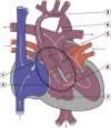
| File:HeartILP draft tetralogyoffallot.jpg | Tetralogy of Fallot | 9-14% of CHD | Classic group of defects: pulmonary stenosis, VSD, dextroposition of aorta, RV hypertrophy. Results in cyanosis. |
| File:HeartILP draft transposition.jpg | Transposition of the Great Vessels | 10-11% of CHD | Aorta arises from the RV with the pulmonary trunk arising from the left. Most common cause of cyanotic heart disease in newborns; surgically corrected. |
| File:HeartILP draft asd.jpg | Atrial Septal Defect | 6-10% of CHD; more common in females | Most commonly patent foramen ovale; can also be an ostium secundum defect, and endocardial cushion defect with ostium primum defect, sinus venosus defect, common atrium. Results in cyanosis due to right-to-left shunt. |
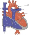
| File:HeartILP draft pulmonaryatresia.jpgFile:HeartILP draft pulmonarystenosis.jpg | Pulmonary Atresia & Pulmonary Stenosis | 10% of CHD | Unequal division of trunks causes cusps to fuse to form a dome with a narrow/non existent lumen. Heart-lung transplantation may be the only therapy |
| File:HeartILP draft patentda.jpg | Patent Ductus Arteriosus | 6-8% of CHD; 2-3 times more common in females; common in preterm newborns | Failure of contraction of the muscular wall of the DA. Spontaneous or surgical closure. |
| File:HeartILP draft aorticstenosis.jpg | Aortic Stenosis | 7% of CHD | Persistence of tissue that normally degenerates. Results in LV hypertrophy, heart murmurs. |
File:HeartILP draft hlh.jpg
|
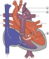
Hypoplastic Left Heart Syndrome | 4-8% of CHD | RV maintains both pulmonary and systemic circulations aided by an ASD. Infants usually die within weeks. |

|
Coarctation of the Aorta | 5-7% of CHD | Aortic constriction. Treatment aims at maintaining the ductus arteriosus via prostaglandins. |
| File:HeartILP draft papvd.jpgFile:HeartILP draft tapvc.jpg | Partial/Total Anomalous Pulmonary Venous Connection | <4% of CHD; more common in females | Total or partial lack of connection of the pulmonary veins with LA. They open into RA, one of the systemic veins or both. The overloaded pulmonary circuit leads to cyanosis, tachypnoea and dyspnoea. Treatment is via surgical redirection |
| File:HeartILP draft tricuspidatresia.jpg | Tricuspid Atresia | 1-3% of CHD | Complete lack of formation of the tricuspid valve with absence of direct connection between the right atrium and right ventricle. Results in cyanosis. |
| File:HeartILP draft dorv.jpg | Double Outlet Right Ventricle | 1-1.5% of CHD | Both great arteries arise wholly or in large part from the right ventricle. Arrangement of the atrioventricular valves and the ventriculoarterial connections are variable. Clinical manifestations variable. |

|
Interrupted Aortic Arch | Very rare | Gap in ascending and descending thoracic aorta. Treated with prostaglandin to maintain ductus arteriosus followed by surgery. |
| Back to Valve Development | Next: Vascular Development | |
| Go to this section in the advanced level |