Integumentary System - Tooth Development: Difference between revisions
mNo edit summary |
mNo edit summary |
||
| Line 26: | Line 26: | ||
| | | | ||
* '''Autocrine regulation of mesenchymal progenitor cell fates orchestrates tooth eruption'''<ref>Akira Takahashi, etal. Autocrine regulation of mesenchymal progenitor cell fates orchestrates tooth eruption. Proceedings of the National Academy of Sciences Jan 2019, 116 (2) 575-580; DOI: 10.1073/pnas.1810200115 https://www.pnas.org/content/116/2/575</ref> "Formation of functional skeletal tissues requires highly organized steps of mesenchymal progenitor cell differentiation. The dental follicle (DF) surrounding the developing tooth harbors mesenchymal progenitor cells for various differentiated cells constituting the tooth root–bone interface and coordinates tooth eruption in a manner dependent on signaling by parathyroid hormone-related peptide (PTHrP) and the PTH/PTHrP receptor (PPR). However, the identity of mesenchymal progenitor cells in the DF and how they are regulated by PTHrP-PPR signaling remain unknown. Here, we show that the PTHrP-PPR autocrine signal maintains physiological cell fates of DF mesenchymal progenitor cells to establish the functional periodontal attachment apparatus and orchestrates tooth eruption. A single-cell RNA-seq analysis revealed cellular heterogeneity of PTHrP+ cells, wherein PTHrP+ DF subpopulations abundantly express PPR. Cell lineage analysis using tamoxifen-inducible PTHrP-creER mice revealed that PTHrP+ DF cells differentiate into cementoblasts on the acellular cementum, periodontal ligament cells, and alveolar cryptal bone osteoblasts during tooth root formation. PPR deficiency induced a cell fate shift of PTHrP+ DF mesenchymal progenitor cells to nonphysiological cementoblast-like cells precociously forming the cellular cementum on the root surface associated with up-regulation of Mef2c and matrix proteins, resulting in loss of the proper periodontal attachment apparatus and primary failure of tooth eruption, closely resembling human genetic conditions caused by PPR mutations." | * '''Autocrine regulation of mesenchymal progenitor cell fates orchestrates tooth eruption'''<ref>Akira Takahashi, etal. Autocrine regulation of mesenchymal progenitor cell fates orchestrates tooth eruption. Proceedings of the National Academy of Sciences Jan 2019, 116 (2) 575-580; DOI: 10.1073/pnas.1810200115 https://www.pnas.org/content/116/2/575</ref> "Formation of functional skeletal tissues requires highly organized steps of mesenchymal progenitor cell differentiation. The dental follicle (DF) surrounding the developing tooth harbors mesenchymal progenitor cells for various differentiated cells constituting the tooth root–bone interface and coordinates tooth eruption in a manner dependent on signaling by parathyroid hormone-related peptide (PTHrP) and the PTH/PTHrP receptor (PPR). However, the identity of mesenchymal progenitor cells in the DF and how they are regulated by PTHrP-PPR signaling remain unknown. Here, we show that the PTHrP-PPR autocrine signal maintains physiological cell fates of DF mesenchymal progenitor cells to establish the functional periodontal attachment apparatus and orchestrates tooth eruption. A single-cell RNA-seq analysis revealed cellular heterogeneity of PTHrP+ cells, wherein PTHrP+ DF subpopulations abundantly express PPR. Cell lineage analysis using tamoxifen-inducible PTHrP-creER mice revealed that PTHrP+ DF cells differentiate into cementoblasts on the acellular cementum, periodontal ligament cells, and alveolar cryptal bone osteoblasts during tooth root formation. PPR deficiency induced a cell fate shift of PTHrP+ DF mesenchymal progenitor cells to nonphysiological cementoblast-like cells precociously forming the cellular cementum on the root surface associated with up-regulation of Mef2c and matrix proteins, resulting in loss of the proper periodontal attachment apparatus and primary failure of tooth eruption, closely resembling human genetic conditions caused by PPR mutations." | ||
* '''Upstream Enhancer Elements of Shh Regulate Oral and Dental Patterning'''{{#pmid:29481312|PMID29481312}} "Sonic hedgehog ( Shh) is important in pattern formation during development. Shh transcription is modulated by a long-range regulatory mechanism containing a number of enhancers, which are spread over nearly 850 kb in the mouse genome. Shh enhancers in the nervous system have been found between intron and 430 kb upstream of Shh. Enhancers in the oral cavity, pharynx, lung, gut, and limbs have been discovered between 610 kb and 850 kb upstream of Shh. However, the intergenic region ranging from 430 to 610 kb upstream of Shh remains to be elucidated. In the present study, we found a novel long-range enhancer located 558 kb upstream of Shh. The enhancer showed in vivo activity in oral cavity and whiskers. A targeted deletion from the novel enhancer to mammal reptile conserved sequence 1 (MRCS1), which is a known enhancer of Shh in oral cavity, resulted in supernumerary molar formation, confirming the essential role of this intergenic region for Shh transcription in teeth. Furthermore, we clarified the binding of Lef1/Tcfs to the new enhancer and MRCS1, suggesting that Wnt/β-catenin signaling regulates Shh signaling in the oral cavity via these enhancers." {{SHH}} | * '''Upstream Enhancer Elements of Shh Regulate Oral and Dental Patterning'''{{#pmid:29481312|PMID29481312}} "Sonic hedgehog ( Shh) is important in pattern formation during development. Shh transcription is modulated by a long-range regulatory mechanism containing a number of enhancers, which are spread over nearly 850 kb in the mouse genome. Shh enhancers in the nervous system have been found between intron and 430 kb upstream of Shh. Enhancers in the oral cavity, pharynx, lung, gut, and limbs have been discovered between 610 kb and 850 kb upstream of Shh. However, the intergenic region ranging from 430 to 610 kb upstream of Shh remains to be elucidated. In the present study, we found a novel long-range enhancer located 558 kb upstream of Shh. The enhancer showed in vivo activity in oral cavity and whiskers. A targeted deletion from the novel enhancer to mammal reptile conserved sequence 1 (MRCS1), which is a known enhancer of Shh in oral cavity, resulted in supernumerary molar formation, confirming the essential role of this intergenic region for Shh transcription in teeth. Furthermore, we clarified the binding of Lef1/Tcfs to the new enhancer and MRCS1, suggesting that Wnt/β-catenin signaling regulates Shh signaling in the oral cavity via these enhancers." {{SHH}} | ||
Revision as of 11:52, 9 January 2019
| Embryology - 25 Apr 2024 |
|---|
| Google Translate - select your language from the list shown below (this will open a new external page) |
|
العربية | català | 中文 | 中國傳統的 | français | Deutsche | עִברִית | हिंदी | bahasa Indonesia | italiano | 日本語 | 한국어 | မြန်မာ | Pilipino | Polskie | português | ਪੰਜਾਬੀ ਦੇ | Română | русский | Español | Swahili | Svensk | ไทย | Türkçe | اردو | ייִדיש | Tiếng Việt These external translations are automated and may not be accurate. (More? About Translations) |
Introduction
The tooth is an extrordinary integumentary system specialization providing insights into epitheilal/mesenchymal (ectoderm of the first pharyngeal arch and neural crest, ectomesenchymal cells) interactions in development and has a major contribution from the neural crest. (More? Neural Crest Development)
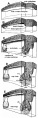
There are 4 morphological stages describing the early tooth development: bud, cap, bell, and terminal differentiation.
| lamina | placode | bud | cap | bell |
|---|---|---|---|---|

|

|

|

|

|
Links: Gastrointestinal Tract Development
| Historic Tooth Development |
|---|
| 1902 Tooth Development | 1912 The Teeth | 1921 The Teeth |
Some Recent Findings

|
| More recent papers |
|---|
 This table allows an automated computer search of the external PubMed database using the listed "Search term" text link.
More? References | Discussion Page | Journal Searches | 2019 References | 2020 References Search term: Tooth Embryology | Tooth Development |
| Older papers |
|---|
| These papers originally appeared in the Some Recent Findings table, but as that list grew in length have now been shuffled down to this collapsible table.
See also the Discussion Page for other references listed by year and References on this current page.
|
Textbooks
- Human Embryology (2nd ed.) Larson Chapter 14 p443-455
- The Developing Human: Clinically Oriented Embryology (6th ed.) Moore and Persaud Chapter 20: P513-529
- Before We Are Born (5th ed.) Moore and Persaud Chapter 21: P481-496
- Essentials of Human Embryology Larson Chapter 14: P303-315
- Human Embryology, Fitzgerald and Fitzgerald
- Color Atlas of Clinical Embryology Moore Persaud and Shiota Chapter 15: p231-236
Movies
|
This time-lapse movie from a mouse embryo (E 12.5–13.5) cultured for 5 days ex vivo, images were taken at 30-min intervals.[7]
The tooth germ is from a developing molar and the lingual side is on the left.
|
Development Overview
- ectoderm, mesoderm and neural crest ectomesenchyme contribute
- inductive influence of neural crest with overlying ectoderm
Odontoblast Cells
The odontoblast cells are a population of neural crest-derived mesenchymal cells.
- differentiate under the influence of the enamel epithelium
- form predentin
- calcifies to form dentin
Ameloblast Cells
The ameloblast cells a population of ectoderm-derived oral epithelium cells that produce the tooth enamal.
- Molecular - BMP and FGF
- tooth growth occurs in ossifying jaws
- periodontal ligament holds tooth in bone socket
Tooth Stages

| Stage | Human (weeks) |
Mouse (days) | |
| lamina |
|
Week 6 | E 11 |
| placode |
|
Week 7 | E 11.5 |
| bud |
|
Week 8 | E 12.5 |
| cap |
|
Week 11 | E 14.5 |
| bell |
|
Week 14 | E 15.5 |
| Tooth Stages | |||
|---|---|---|---|
| Stage | Human (weeks) |
Mouse (days) | |
| lamina |
|
Week 6 | E11 |
| placode |
|
Week 7 | E11.5 |
| bud |
|
Week 8 | E12.5 |
| cap |
|
Week 11 | E14.5 |
| bell |
|
Week 14 | E15.5 |
Image Links: all stages | lamina | placode stage | bud stage | cap stage | bell stage
Human 2 Sets of Teeth

Human Dentition Timeline
| The milk dentition | |
|---|---|
| Median incisors | 6th to 8th month |
| Lateral incisors | 8th to 12th month |
| First molars | 12th to 16th month |
| Canines 1 | 7th to 20th month |
| Second molars | 20th to 24th month |
| The permanent dentition | |
| First molars | 7th year |
| Median incisors | 8th year |
| Lateral incisors | 9th year |
| First premolars | 10th year |
| Second premolars | 11th year |
| Canines | 13th to 14th year |
| Second molars | 13th to 14th year |
| Third molars | 17th to 40th year |
- Source: The Teeth (1912)[9]
Deciduous Teeth
- 20 deciduous teeth
- Differential rates of growth, shed at different times over 20 year period

Permanent Teeth

- 32 permanent teeth
- Incisors - sharp cutting edge, adapted for biting the food.
- Canines - are larger and stronger than the incisors. The upper canines have also been called the "eye teeth", while the lower canines "stomach teeth".
- Premolars - or Bicuspid teeth are smaller and shorter than the canines.
- Molars - are the largest teeth adapted for grinding and pounding food.

Epithelial Mesenchymal Interaction
local ectodermal thickening expresses several signaling molecules these in turn signal to the underlying mesenchyme triggering mesenchymal condensation (epithelially expressed Bmp4 induces Msx1 and Lef1 as well as itself in the underlying mesenchyme)
Four epithelial signaling molecules, Bmp2, Shh, Wnt10a, and Wnt10b, in the early inductive cascade, each signal has a distinct molecular action on the jaw mesenchyme.
Mouse (E11.0 and E12.0) - all four genes are specifically expressed in the epithelium.
Shh and Wnt10b induce general Hedgehog and Wnt targets, Ptc and Gli for Shh and Lef1 for Wnt10b,
Bmp2 is able to induce tooth-specific expression of Msx1.
(Text above modified from: Hélène R. Dassule and Andrew P. McMahon Developmental Biology, v 202, n 2, October 15, 1998, p215-227)
(More? Developmental Mechanism - Epithelial Mesenchymal Interaction)
Periodontal Ligament
The tooth is not anchored directly onto its bony socket (alveolar bone) but held in place by the periodontal ligament (PDL), a specialized connective tissue structure that surrounds the tooth root coating of cementum.
The additional roles of the PDL are to also act as; a shock absorber, transmitter of chewing forces (from tooth to bone), sensory information (heat, cold, pressure and pain).
The collagen fiber bundles within the ligament are called "Sharpey’s fibres".
Cementum (from investing layer of the dental follicle) is contiguous layer with the periodontal ligament on one surface and firmly adherent to dentine on the other surface.
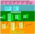
Molecular Tooth Development

More than 300 genes have been associated with tooth development including: BMP4, FGF8, MSX1, MSX2, PAX9, PITX2, SHOX2, Delta/Notch, Hox-8, Runx2
Most recent review in Developmental Dynamics by Lin D, Huang Y, He F, Gu S, Zhang G, Chen Y, Zhang Y. Expression survey of genes critical for tooth development in the human embryonic tooth germ. Dev Dyn. 2007 Mar 29.
Amelogenin - abundant protein secreted by ameloblasts which is a major component of tooth enamel.
The papers below are from UNSW Embryology (version 3), information requires updating.
Bone Morphogenic Protein (BMP) / Fibroblast Growth Factor (FGF)
Growth factors in the BMP- and FGF-families are expressed in dental epithelium during initiation of tooth development and their effects on the underlying mesenchyme mimic those of the epithelium. They upregulate the expression of many genes, including the homeobox-containing Msx-1 and Msx-2, and stimulate cell proliferation suggesting that they may act as epithelial signals transmitting epithelial-mesenchymal interactions. During subsequent morphogenesis, when the characteristic shapes of individual teeth develop as a result from folding of the dental epithelium, several signal molecules including Sonic hedgehog, Bmps-2, 4, 7 and Fgf-4 are expressed specifically in restricted and transient epithelial cell clusters, called enamel knots.
(Text: Irma Thesleff and Carin Sahlberg Seminars in Cell & Developmental Biology, v 7, n 2, April, 1996, p185-193)
Delta/Notch
The expression pattern of Delta 1 in ameloblasts and odontoblasts is complementary to Notch1, Notch2, and Notch3 expression in adjacent epithelial and mesenchymal cells. Notch1 and Notch2 are upregulated in explants of dental mesenchyme adjacent to implanted cells expressing Delta1, suggesting that feedback regulation by Delta-Notch signaling ensures the spatial segregation of Notch receptors and ligands. TGF1 and BMPs induce Delta1 expression in dental mesenchyme explants at the stage at which Delta1 is upregulated in vivo, but not at earlier stages. In contrast to the Notch family receptors and their ligand Jagged1, expression of Delta1 in the tooth germ is not affected by epithelial-mesenchymal interactions, showing that the Notch receptors and their two ligands Jagged1 and Delta1 are subject to different regulations.
Text: Mitsiadis etal Developmental Biology,v 204, n 2, December 15, 1998, p420-431
BMP4 expression by the interaction of Pax9 with Msx1 at the level of transcription and protein complex determines the fate of the transition from bud to cap stage during tooth development.[10]
Twist1 a basic helix-loop-helix-containing transcription factor expressed in dental mesenchyme during the early stages of tooth development acts through the FGF signaling pathway.[11]
Foxi3 forkhead-box transcription factor inhibits formation of enamel knots and cervical loops and therefore the differentiation of dental epithelium.[12]
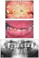
Abnormalities

Adontia
A total lack of tooth development.
Dentinogenesis imperfecta
The teeth are translucent and often roughened with severe amber discolouration. Discoloured teeth with an opalescent sheen, dentin does not support enamel (dentin sialophosphoprotein mutation)
Dentine dysplasia
The primary teeth are translucent and amber in colour whereas the erupting secondary central incisors are of normal appearance.
Amelogenesis Imperfecta
Abnormal tooth enamel formation (AMELX, ENAM, KLK4, MMP20).
Dens Evaginatus
Dental anomaly mainly affecting premolars in people of Mongolian origin.
Hypodontia
Lack of development of one or more teeth.
Hypohidrotic Ectodermal Dysplasia
Maldevelopment of one or more ectodermal-derived tissues.
Microdontia
Small teeth.
Hutchinson's teeth
(Hutchinson's incisor, Hutchinson's sign, Hutchinson-Boeck teeth) Historic clinical term for an infant tooth abnormality associated with congenital syphilis. Teeth are smaller, more widely spaced than normal and have notches on the biting surfaces. Named after Jonathan Hutchinson (1828 – 1913) an English surgeon and pathologist, who first described this association.
References
- ↑ Akira Takahashi, etal. Autocrine regulation of mesenchymal progenitor cell fates orchestrates tooth eruption. Proceedings of the National Academy of Sciences Jan 2019, 116 (2) 575-580; DOI: 10.1073/pnas.1810200115 https://www.pnas.org/content/116/2/575
- ↑ Seo H, Amano T, Seki R, Sagai T, Kim J, Cho SW & Shiroishi T. (2018). Upstream Enhancer Elements of Shh Regulate Oral and Dental Patterning. J. Dent. Res. , , 22034518758642. PMID: 29481312 DOI.
- ↑ Bonczek O, Balcar VJ & Šerý O. (2017). PAX9 gene mutations and tooth agenesis: A review. Clin. Genet. , 92, 467-476. PMID: 28155232 DOI.
- ↑ Li J, Chatzeli L, Panousopoulou E, Tucker AS & Green JB. (2016). Epithelial stratification and placode invagination are separable functions in early morphogenesis of the molar tooth. Development , 143, 670-81. PMID: 26755699 DOI.
- ↑ Huang X, Bringas P, Slavkin HC & Chai Y. (2009). Fate of HERS during tooth root development. Dev. Biol. , 334, 22-30. PMID: 19576204 DOI.
- ↑ Lin D, Huang Y, He F, Gu S, Zhang G, Chen Y & Zhang Y. (2007). Expression survey of genes critical for tooth development in the human embryonic tooth germ. Dev. Dyn. , 236, 1307-12. PMID: 17394220 DOI.
- ↑ Morita R, Kihira M, Nakatsu Y, Nomoto Y, Ogawa M, Ohashi K, Mizuno K, Tachikawa T, Ishimoto Y, Morishita Y & Tsuji T. (2016). Coordination of Cellular Dynamics Contributes to Tooth Epithelium Deformations. PLoS ONE , 11, e0161336. PMID: 27588418 DOI.
- ↑ 8.0 8.1 Koussoulakou DS, Margaritis LH & Koussoulakos SL. (2009). A curriculum vitae of teeth: evolution, generation, regeneration. Int. J. Biol. Sci. , 5, 226-43. PMID: 19266065
- ↑ Keibel F. and Mall FP. Manual of Human Embryology II. (1912) J. B. Lippincott Company, Philadelphia.
- ↑ Ogawa T, Kapadia H, Feng JQ, Raghow R, Peters H & D'Souza RN. (2006). Functional consequences of interactions between Pax9 and Msx1 genes in normal and abnormal tooth development. J. Biol. Chem. , 281, 18363-9. PMID: 16651263 DOI.
- ↑ Meng T, Huang Y, Wang S, Zhang H, Dechow PC, Wang X, Qin C, Shi B, D'Souza RN & Lu Y. (2015). Twist1 Is Essential for Tooth Morphogenesis and Odontoblast Differentiation. J. Biol. Chem. , 290, 29593-602. PMID: 26487719 DOI.
- ↑ Jussila M, Aalto AJ, Sanz Navarro M, Shirokova V, Balic A, Kallonen A, Ohyama T, Groves AK, Mikkola ML & Thesleff I. (2015). Suppression of epithelial differentiation by Foxi3 is essential for molar crown patterning. Development , 142, 3954-63. PMID: 26450968 DOI.
- ↑ Barron MJ, McDonnell ST, Mackie I & Dixon MJ. (2008). Hereditary dentine disorders: dentinogenesis imperfecta and dentine dysplasia. Orphanet J Rare Dis , 3, 31. PMID: 19021896 DOI.
Journals
Reviews
Peterkova R, Hovorakova M, Peterka M & Lesot H. (2014). Three-dimensional analysis of the early development of the dentition. Aust Dent J , 59 Suppl 1, 55-80. PMID: 24495023 DOI.
Bei M. (2009). Molecular genetics of tooth development. Curr. Opin. Genet. Dev. , 19, 504-10. PMID: 19875280 DOI.
Seppala M, Zoupa M, Onyekwelu O & Cobourne MT. (2006). Tooth development: 1. Generating teeth in the embryo. Dent Update , 33, 582-4, 586-8, 590-1. PMID: 17209531
Thesleff I. (2006). The genetic basis of tooth development and dental defects. Am. J. Med. Genet. A , 140, 2530-5. PMID: 16838332 DOI.
Tompkins K. (2006). Molecular mechanisms of cytodifferentiation in mammalian tooth development. Connect. Tissue Res. , 47, 111-8. PMID: 16753804 DOI.
Cobourne MT & Sharpe PT. (2003). Tooth and jaw: molecular mechanisms of patterning in the first branchial arch. Arch. Oral Biol. , 48, 1-14. PMID: 12615136
Sharpe PT. (2001). Neural crest and tooth morphogenesis. Adv. Dent. Res. , 15, 4-7. PMID: 12640730 DOI.
Articles
Lin D, Huang Y, He F, Gu S, Zhang G, Chen Y & Zhang Y. (2007). Expression survey of genes critical for tooth development in the human embryonic tooth germ. Dev. Dyn. , 236, 1307-12. PMID: 17394220 DOI.
Nakatomi M, Morita I, Eto K & Ota MS. (2006). Sonic hedgehog signaling is important in tooth root development. J. Dent. Res. , 85, 427-31. PMID: 16632755 DOI.
Ogawa T, Kapadia H, Feng JQ, Raghow R, Peters H & D'Souza RN. (2006). Functional consequences of interactions between Pax9 and Msx1 genes in normal and abnormal tooth development. J. Biol. Chem. , 281, 18363-9. PMID: 16651263 DOI.
Kettunen P & Thesleff I. (1998). Expression and function of FGFs-4, -8, and -9 suggest functional redundancy and repetitive use as epithelial signals during tooth morphogenesis. Dev. Dyn. , 211, 256-68. PMID: 9520113 <256::AID-AJA7>3.0.CO;2-G DOI.
Search PubMed
Search Pubmed: Tooth Development | odontogenesis | tooth morphogenesis | adontia | amelogenesis imperfecta | dens evaginatus | hypodontia
Additional Images
Category:Tooth | Category:Integumentary
Tooth development stage
Tooth molecular development
Inherited dentine disorders
Mouse - lower incisor teeth
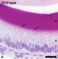
Mouse - tooth histology
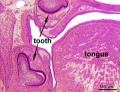
Rat - neonatal teeth


Historic
Development and Morphology of the Teeth (1902)
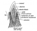
Fig. 47. Showing the parts of an incisor tooth.
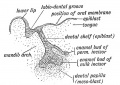
Fig. 48. Section through the lip and mandible of a foetus in the third month, showing the down-growth of the Dental Shelf.
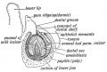
Fig. 49. Showing the stage of development in an incisor tooth of a foetus of six months.
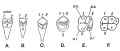
Fig. 50. A. The tritubercular Type of Tooth.


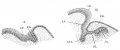
Fig. 262. Section through the dental ridge of the lower jaw of embryos.
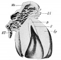
Fig. 263. Reconstruction of the dental ridge and a papilla of an embryo of 30 cm.
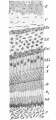
Fig. 264. Section through a developing molar tooth of Didelphys
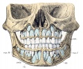
Fig. 265. Skull of a 5-year-old child showing the milk and permanent dentitions
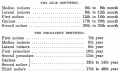
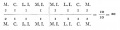
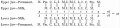
Table - Dentition Timeline


Fig. 252. Section of developing tooth from a 3 months human fetus.
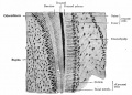
Fig. 253. Section through the border of a developing tooth of a new-born puppy.
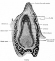
Fig. 254. Longitudinal section of a developing tooth of a new-born puppy
Milk teeth
Permanent teeth


Terms
External Links
External Links Notice - The dynamic nature of the internet may mean that some of these listed links may no longer function. If the link no longer works search the web with the link text or name. Links to any external commercial sites are provided for information purposes only and should never be considered an endorsement. UNSW Embryology is provided as an educational resource with no clinical information or commercial affiliation.
- StemBook - Tooth organogenesis and regeneration
- University of Helsinki Gene Expression in Tooth
- American Dental Association Overview - Tooth
- Columbia University Medical Centre Illustrations: How a Tooth Decays
- Merck Tooth disorders
- Nemours Foundation Teething Tots
Glossary Links
- Glossary: A | B | C | D | E | F | G | H | I | J | K | L | M | N | O | P | Q | R | S | T | U | V | W | X | Y | Z | Numbers | Symbols | Term Link
Cite this page: Hill, M.A. (2024, April 25) Embryology Integumentary System - Tooth Development. Retrieved from https://embryology.med.unsw.edu.au/embryology/index.php/Integumentary_System_-_Tooth_Development
- © Dr Mark Hill 2024, UNSW Embryology ISBN: 978 0 7334 2609 4 - UNSW CRICOS Provider Code No. 00098G