Integumentary System - Eyelid Development: Difference between revisions
mNo edit summary |
mNo edit summary |
||
| Line 111: | Line 111: | ||
File:Gray0897.jpg|897 Structures of the Lacrimal Gland | File:Gray0897.jpg|897 Structures of the Lacrimal Gland | ||
</gallery> | </gallery> | ||
==External Links== | |||
{{External Links}} | |||
* [https://www.best.edu.au/s/share?url=ckjlb65f%2Fwfuc4g5f&data=8%401!9%4027360!10%40-12267&version=1 Histology - Adult Eyelid] | |||
{{Glossary}} | {{Glossary}} | ||
Revision as of 21:48, 8 January 2019
Introduction
The eyelids represent integumentary specialisations that cover and protect the eye cornea.
Note that some species, such as rodents, are born with closed eyelids.
The palpebral commissure (canthus) is located at the corner of the eye where the upper and lower eyelids meet.


| Vision Links: vision | lens | retina | placode | extraocular muscle | cornea | eyelid | lacrima gland | vision abnormalities | Student project 1 | Student project 2 | Category:Vision | sensory | ||
|
Some Recent Findings
|
| More recent papers |
|---|
 This table allows an automated computer search of the external PubMed database using the listed "Search term" text link.
More? References | Discussion Page | Journal Searches | 2019 References | 2020 References Search term: Eyelid Embryology | Eyelid Development |
| Older papers |
|---|
| These papers originally appeared in the Some Recent Findings table, but as that list grew in length have now been shuffled down to this collapsible table.
See also the Discussion Page for other references listed by year and References on this current page.
|
Human Eyelid Timeline
| Carnegie Stage | Event |
|---|---|
| 10 | optic primordia appear |
| 13 | By the end of the fourth week the optic vesicle lies close to the surface ectoderm. The surface ectoderm overlying the optic vesicle, in response to this contact, has thickened to form the lense placode. |
| 14 | (about 32 days) the lens placode is indented by the lens pit. |
| 15 | (about 33 days) the lens pit is closed. The lens vesicle and optic cup lie close to the surface ectoderm and appear to press against the surface. |
| 16 | (37 days) Prior to the development of the eyelids, one small sulcus or groove forms above the eye (eyelid groove) and another below it. |
| 17 - 19 | grooves deepen, eyelid folds develop, first below, and then above, the eye. |
| 19 - 22 | eyelid folds develop into the eyelids and cover more of the eye as the palpebral fissure takes shape. The upper and the lower eyelids meet at the outer canthus in Stage 19. |
| 20 | the inner canthus is established. |
| 23 | closure of the eyelids is complete (Note - shown as still open in some Kyoto embryo). |
| Data - from Kyoto embryos with Carnegie staging.[5] Links: eyelid |
The images below link to virtual slides of the human developing eye at Carnegie stage 22. Click on the image to open or select specific regions from the regions of interest links.
|
|
Virtual Slide - Regions of Interest
Links: Embryo Virtual Slides |


Adult Anatomy

The palpebral commissure (canthus) is located at the corner of the eye where the upper and lower eyelids meet.
Molecular
See recent article on molecular biology and genetics of embryonic eyelid development.[1]
- cell migration - FGF10, TGF-α, Activin B, and HB-EGF modulate downstream BMP4 signaling, the ERK cascade, and JNK/c-JUN.
- Wnt/β-catenin signaling pathway - may inhibit and regulate eyelid fusion.
Abnormalities
Congenital upper eyelid ectopic cilia
References
- ↑ 1.0 1.1 Rubinstein TJ, Weber AC & Traboulsi EI. (2016). Molecular biology and genetics of embryonic eyelid development. Ophthalmic Genet. , 37, 252-9. PMID: 26863902 DOI.
- ↑ Sanchis A, Bayo P, Sevilla LM & Pérez P. (2010). Glucocorticoid receptor antagonizes EGFR function to regulate eyelid development. Int. J. Dev. Biol. , 54, 1473-80. PMID: 21136383 DOI.
- ↑ Mine N, Iwamoto R & Mekada E. (2005). HB-EGF promotes epithelial cell migration in eyelid development. Development , 132, 4317-26. PMID: 16141218 DOI.
- ↑ Zhang H, Hara M, Seki K, Fukuda K & Nishida T. (2005). Eyelid fusion and epithelial differentiation at the ocular surface during mouse embryonic development. Jpn. J. Ophthalmol. , 49, 195-204. PMID: 15944823 DOI.
- ↑ Pearson AA. (1980). The development of the eyelids. Part I. External features. J. Anat. , 130, 33-42. PMID: 7364662
Reviews
Zieske JD. (2004). Corneal development associated with eyelid opening. Int. J. Dev. Biol. , 48, 903-11. PMID: 15558481 DOI.
Hamming N. (1983). Anatomy and embryology of the eyelids: a review with special reference to the development of divided nevi. Pediatr Dermatol , 1, 51-8. PMID: 6387662
Articles
Meng Q, Mongan M, Carreira V, Kurita H, Liu CY, Kao WW & Xia Y. (2014). Eyelid closure in embryogenesis is required for ocular adnexa development. Invest. Ophthalmol. Vis. Sci. , 55, 7652-61. PMID: 25377219 DOI.
Du X, Tabeta K, Hoebe K, Liu H, Mann N, Mudd S, Crozat K, Sovath S, Gong X & Beutler B. (2004). Velvet, a dominant Egfr mutation that causes wavy hair and defective eyelid development in mice. Genetics , 166, 331-40. PMID: 15020428
Findlater GS, McDougall RD & Kaufman MH. (1993). Eyelid development, fusion and subsequent reopening in the mouse. J. Anat. , 183 ( Pt 1), 121-9. PMID: 8270467
Harris MJ & McLeod MJ. (1982). Eyelid growth and fusion in fetal mice. A scanning electron microscope study. Anat. Embryol. , 164, 207-20. PMID: 7125235
Search PubMed
Search Pubmed: Eyelid Development
Additional Images
Historic
| Historic Disclaimer - information about historic embryology pages |
|---|
|
Gray H. Anatomy of the human body. (1918) Philadelphia: Lea & Febiger.
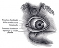
892 Front of left eye with eyelids separated to show medial canthus
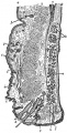
893 Structure of the Eyelids
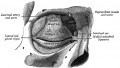
894 The tarsi and their ligaments
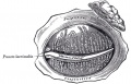
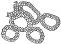
895 The Tarsal Glands
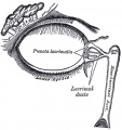
896 The Lacrimal Gland
897 Structures of the Lacrimal Gland


External Links
External Links Notice - The dynamic nature of the internet may mean that some of these listed links may no longer function. If the link no longer works search the web with the link text or name. Links to any external commercial sites are provided for information purposes only and should never be considered an endorsement. UNSW Embryology is provided as an educational resource with no clinical information or commercial affiliation.
Glossary Links
- Glossary: A | B | C | D | E | F | G | H | I | J | K | L | M | N | O | P | Q | R | S | T | U | V | W | X | Y | Z | Numbers | Symbols | Term Link
Cite this page: Hill, M.A. (2024, April 19) Embryology Integumentary System - Eyelid Development. Retrieved from https://embryology.med.unsw.edu.au/embryology/index.php/Integumentary_System_-_Eyelid_Development
- © Dr Mark Hill 2024, UNSW Embryology ISBN: 978 0 7334 2609 4 - UNSW CRICOS Provider Code No. 00098G