Human Development Movie: Difference between revisions
mNo edit summary |
mNo edit summary |
||
| Line 1: | Line 1: | ||
{{Movie header}} | {{Movie header}} | ||
==Human Development Cartoon== | |||
{| | {| | ||
| <mediaplayer width='360' height='410' image="http://embryology.med.unsw.edu.au/embryology/images/6/61/Human_development_001_icon.jpg">File:Human development 001.mp4</mediaplayer> | | <mediaplayer width='360' height='410' image="http://embryology.med.unsw.edu.au/embryology/images/6/61/Human_development_001_icon.jpg">File:Human development 001.mp4</mediaplayer> | ||
| valign=top| | | valign=top|This animation overview begins at the zygote stage following fertilization and takes you through an overview of the entire 9 months of human development in just over a minute! | ||
This animation overview begins at the zygote stage following fertilization and takes you through an overview of the entire 9 months of human development in just over a minute! | |||
| Line 18: | Line 15: | ||
{{Human Development cartoon}} | {{Human Development cartoon}} | ||
|} | |||
| | ==Events in this Animation== | ||
{| | |||
| <mediaplayer width='360' height='410' image="http://embryology.med.unsw.edu.au/embryology/images/6/61/Human_development_001_icon.jpg">File:Human development 001.mp4</mediaplayer> | | <mediaplayer width='360' height='410' image="http://embryology.med.unsw.edu.au/embryology/images/6/61/Human_development_001_icon.jpg">File:Human development 001.mp4</mediaplayer> | ||
| | | | ||
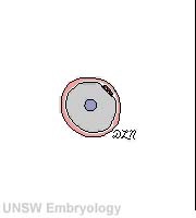
'''00:02''' - Two [[B#blastomere|blastomere]] cell stage: enclosed within the [[Z#zona pellucida|zona pellucida]] (pink ring) with [[P#polar body|polar bodies]] (upper right) lying under the zona pellucida. | '''00:02''' - Two [[B#blastomere|blastomere]] cell stage: enclosed within the [[Z#zona pellucida|zona pellucida]] (pink ring) with [[P#polar body|polar bodies]] (upper right) lying under the zona pellucida. | ||
Revision as of 10:57, 15 April 2013
| Embryology - 16 Apr 2024 |
|---|
| Google Translate - select your language from the list shown below (this will open a new external page) |
|
العربية | català | 中文 | 中國傳統的 | français | Deutsche | עִברִית | हिंदी | bahasa Indonesia | italiano | 日本語 | 한국어 | မြန်မာ | Pilipino | Polskie | português | ਪੰਜਾਬੀ ਦੇ | Română | русский | Español | Swahili | Svensk | ไทย | Türkçe | اردو | ייִדיש | Tiếng Việt These external translations are automated and may not be accurate. (More? About Translations) |
Human Development Cartoon
| <mediaplayer width='360' height='410' image="http://embryology.med.unsw.edu.au/embryology/images/6/61/Human_development_001_icon.jpg">File:Human development 001.mp4</mediaplayer> | This animation overview begins at the zygote stage following fertilization and takes you through an overview of the entire 9 months of human development in just over a minute!
|
Events in this Animation
| <mediaplayer width='360' height='410' image="http://embryology.med.unsw.edu.au/embryology/images/6/61/Human_development_001_icon.jpg">File:Human development 001.mp4</mediaplayer> |
00:02 - Two blastomere cell stage: enclosed within the zona pellucida (pink ring) with polar bodies (upper right) lying under the zona pellucida. 00:06 - Morula stage: a solid ball of cells, still dividing rapidly. 00:08 - Blastocyst stage: trophoblast cells (at periphery), embryoblast (inner cell mass, right) and blastocoel (fluid-filled cavity, yellow) still within the zona pellucida. 00:09 - Blastocyst hatches from zona pellucida and adplants (attaches, adheres) to the uterine endometrium epithelium (right red structure) 00:10 - Bilaminar embryo stage: Embryoblast has formed two layers, hypoblast layer (yellow) and epiblast layer (blue). Amniotic cavity (fluid-filled space, blue beside the epiblast) forms, implantation continues into the uterine wall and the trophoblast layer now also forms two cellular layers (outer synctiotrophoblast and inner cytotrophoblast). 00:12 - Conceptus is completely implanted in uterine wall, maternal lacunae (blood-filled spaces, right, dark red) provide nutrition and hCG hormone exchange into the maternal blood. 00:14 - Chorionic cavity (extraembryonic coelom, pale blue space) forms and epiblast gastrulation continues. 00:15 - Yolk sac (yellow cavity, left) formed, now 3 cavities lie outside the embryo. 00:23 - Embryo (dark blue vertical) forms in amniotic cavity and connecting stalk (dark brown, bottom) is the region of eventual placental cord vessel development. Trophoblast layers and extraembryonic mesoderm are forming the early placental villi. 00:24 - Embryo folding with the heart bulge and pharyngeal arches. 00:28 - Embryo growth with limb bud development. 00:38 - Amniotic sac expands and fuses with chorionic cavity wall and the yolk sac remnant is also pushed to the periphery. 00:40 - Week 8, end of the embryonic period of development. 00:41 - Fetal Development from this point onward extensive growth in size occurs during the second and third trimesters. |
Glossary Links: A | B | C | D | E | F | G | H | I | J | K | L | M | N | O | P | Q | R | S | T | U | V | W | X | Y | Z | Numbers | Symbols | Movies
Cite this page: Hill, M.A. (2024, April 16) Embryology Human Development Movie. Retrieved from https://embryology.med.unsw.edu.au/embryology/index.php/Human_Development_Movie
- © Dr Mark Hill 2024, UNSW Embryology ISBN: 978 0 7334 2609 4 - UNSW CRICOS Provider Code No. 00098G