Horse Development: Difference between revisions
mNo edit summary |
mNo edit summary |
||
| Line 66: | Line 66: | ||
==Morula and Blastocyst== | ==Morula and Blastocyst== | ||
The following images are from a historic (1945) article on early horse<ref>Hamilton WJ. [[Paper - Cleavage Stages of the Ova of the Horse with Notes on Ovulation|Cleavage Stages of the Ova of the Horse, with Notes on Ovulation]]. J Anat. 1945 Jul; 79(Pt 3): 127–130.3.</ref> | |||
:[[Paper - Cleavage Stages of the Ova of the Horse with Notes on Ovulation|'''Links''']]: [[:File:Hamilton1945-fig01.jpg|Fig. 1]] | [[:File:Hamilton1945-fig02.jpg|Fig. 2]] | [[:File:Hamilton1945-fig03.jpg|Fig. 3]] | [[:File:Hamilton1945-fig04.jpg|Fig. 4]] | [[:File:Hamilton1945-fig05.jpg|Fig. 5]] | [[:File:Hamilton1945-plate01.jpg|Plate 1]] | [[:File:Hamilton1945-fig06.jpg|Fig 6]] | [[:File:Hamilton1945-fig07.jpg|Fig 7]] | [[:File:Hamilton1945-fig08.jpg|Fig 8]] | [[:File:Hamilton1945-fig09.jpg|Fig 9]] | [[:File:Hamilton1945-fig10.jpg|Fig 10]] | [[:File:Hamilton1945-fig11.jpg|Fig 11]] | [[:File:Hamilton1945-plate02.jpg|Plate 2]] | [[:File:Hamilton1945-fig12.jpg|Fig 12]] | [[:File:Hamilton1945-fig13.jpg|Fig 13]] | [[:File:Hamilton1945-fig14.jpg|Fig 14]] | [[:File:Hamilton1945-fig15.jpg|Fig 15]] | [[:File:Hamilton1945-plate03.jpg|Plate 3]] | |||
{{Historic Disclaimer}} | |||
===Plate 1=== | |||
[[File:Hamilton1945-plate01.jpg|600px]] | |||
<gallery> | |||
File:Hamilton1945-fig01.jpg|Fig. 1. Photograph of a living unsegmented ovum of the pony (P 1) in Locke’s fluid. Granular material is seen in the perivitelline space. X 480. | |||
File:Hamilton1945-fig02.jpg|Fig. 2: Photograph of an unsegmented ovum of the pony (P 1) in agar. The nucleus is just visible. x 480. | |||
File:Hamilton1945-fig03.jpg|Fig. 3. Section of an unsegmented ovum of the pony (P1) with a large vesicular nucleus. x 520. | |||
File:Hamilton1945-fig04.jpg|Fig. 4. Section of an unsegmented ovum of the pony (P 8) showing deutoplasmolysis. x 520. | |||
File:Hamilton1945-fig05.jpg|Fig. 5. Photograph of a living two-cell stage ovum of the pony (P 5). Granular material is seen in the lower part of the perivitelline space. x 480. | |||
</gallery> | |||
===Plate 2=== | |||
[[File:Hamilton1945-plate02.jpg|600px]] | |||
<gallery> | |||
File:Hamilton1945-fig06.jpg|Figs. 6, 7. Two consecutive sections through the two-cell ovum shown in fig. 5. x 520. | |||
File:Hamilton1945-fig07.jpg|Figs. 6, 7. Two consecutive sections through the two-cell ovum shown in fig. 5. x 520. | |||
File:Hamilton1945-fig08.jpg|Fig. 8. Photograph of a living four-cell stage ovum of the pony (P 7). x 480. | |||
File:Hamilton1945-fig09.jpg|Figs. 9, 10. Two consecutive sections through the four-cell ovum shown in fig. 8. x 520. | |||
File:Hamilton1945-fig10.jpg|Figs. 9, 10. Two consecutive sections through the four-cell ovum shown in fig. 8. x 520. | |||
File:Hamilton1945-fig11.jpg|Fig. 11. Photograph of a five-cell stage ovum of the pony (P 4) in agar. x 480. | |||
</gallery> | |||
===Plate 3=== | |||
[[File:Hamilton1945-plate03.jpg|600px]] | |||
<gallery> | |||
File:Hamilton1945-fig12.jpg|Fig. 12. Section through the five-cell ovum shown in fig. 11. x 520. | |||
File:Hamilton1945-fig13.jpg|Fig. 13. Photograph of the living morula. of the pony (P 6). it shows extensive deutoplasmolysis. x 480. | |||
File:Hamilton1945-fig14.jpg|Figs. 14, 15. Two consecutive sections through the morula. shown in fig. 13. x 520. | |||
File:Hamilton1945-fig15.jpg|Figs. 14, 15. Two consecutive sections through the morula. shown in fig. 13. x 520. | |||
</gallery> | |||
Revision as of 12:07, 12 January 2016
| Embryology - 19 Apr 2024 |
|---|
| Google Translate - select your language from the list shown below (this will open a new external page) |
|
العربية | català | 中文 | 中國傳統的 | français | Deutsche | עִברִית | हिंदी | bahasa Indonesia | italiano | 日本語 | 한국어 | မြန်မာ | Pilipino | Polskie | português | ਪੰਜਾਬੀ ਦੇ | Română | русский | Español | Swahili | Svensk | ไทย | Türkçe | اردو | ייִדיש | Tiếng Việt These external translations are automated and may not be accurate. (More? About Translations) |
Introduction
Equine development (Latin, equus = "horse")
| Horse Links: horse | Category:Horse |
| Historic Papers: 1897 Critical Period in Horse Development | 1925 Organ of Jacobson | 1945 Cleavage Stages of the Horse Ova |
| Animal Development: axolotl | bat | cat | chicken | cow | dog | dolphin | echidna | fly | frog | goat | grasshopper | guinea pig | hamster | horse | kangaroo | koala | lizard | medaka | mouse | opossum | pig | platypus | rabbit | rat | salamander | sea squirt | sea urchin | sheep | worm | zebrafish | life cycles | development timetable | development models | K12 |
Some Recent Findings
|
| More recent papers |
|---|
 This table allows an automated computer search of the external PubMed database using the listed "Search term" text link.
More? References | Discussion Page | Journal Searches | 2019 References | 2020 References Search term: Equine Embryology <pubmed limit=5>Equine Embryology</pubmed> |
Taxon
Equine Development
338 days
| Animal Development Time | ||||||||||||||||||||||||||||||||||||||||||||||||||||||||||||||||||||||||||||||||||||||||||||||||||||||||||||||||||||||||||||||||||||||||||||||
|---|---|---|---|---|---|---|---|---|---|---|---|---|---|---|---|---|---|---|---|---|---|---|---|---|---|---|---|---|---|---|---|---|---|---|---|---|---|---|---|---|---|---|---|---|---|---|---|---|---|---|---|---|---|---|---|---|---|---|---|---|---|---|---|---|---|---|---|---|---|---|---|---|---|---|---|---|---|---|---|---|---|---|---|---|---|---|---|---|---|---|---|---|---|---|---|---|---|---|---|---|---|---|---|---|---|---|---|---|---|---|---|---|---|---|---|---|---|---|---|---|---|---|---|---|---|---|---|---|---|---|---|---|---|---|---|---|---|---|---|---|---|---|
| ||||||||||||||||||||||||||||||||||||||||||||||||||||||||||||||||||||||||||||||||||||||||||||||||||||||||||||||||||||||||||||||||||||||||||||||
Animal Notes and Table Data Sources
|
Genetics
Chromosomes: 1 | 2 | 3 | 4 | 5 | 6 | 7 | 8 | 9 | 10 | 11 | 12 |
31 | X | 11 12 13 14 15 16 17 18 19 20 21 22 23 24 25 26 27 28 29 30 31 X
Genome: Equus caballus
Morula and Blastocyst
The following images are from a historic (1945) article on early horse[2]
- Links: Fig. 1 | Fig. 2 | Fig. 3 | Fig. 4 | Fig. 5 | Plate 1 | Fig 6 | Fig 7 | Fig 8 | Fig 9 | Fig 10 | Fig 11 | Plate 2 | Fig 12 | Fig 13 | Fig 14 | Fig 15 | Plate 3
| Historic Disclaimer - information about historic embryology pages |
|---|
|
Plate 1

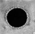
Fig. 1. Photograph of a living unsegmented ovum of the pony (P 1) in Locke’s fluid. Granular material is seen in the perivitelline space. X 480.
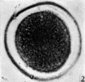
Fig. 2: Photograph of an unsegmented ovum of the pony (P 1) in agar. The nucleus is just visible. x 480.
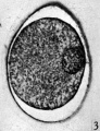
Fig. 3. Section of an unsegmented ovum of the pony (P1) with a large vesicular nucleus. x 520.
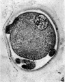
Fig. 4. Section of an unsegmented ovum of the pony (P 8) showing deutoplasmolysis. x 520.
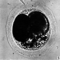
Fig. 5. Photograph of a living two-cell stage ovum of the pony (P 5). Granular material is seen in the lower part of the perivitelline space. x 480.


Plate 2

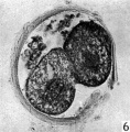
Figs. 6, 7. Two consecutive sections through the two-cell ovum shown in fig. 5. x 520.
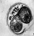
Figs. 6, 7. Two consecutive sections through the two-cell ovum shown in fig. 5. x 520.
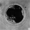
Fig. 8. Photograph of a living four-cell stage ovum of the pony (P 7). x 480.
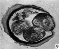
Figs. 9, 10. Two consecutive sections through the four-cell ovum shown in fig. 8. x 520.
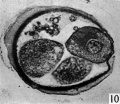
Figs. 9, 10. Two consecutive sections through the four-cell ovum shown in fig. 8. x 520.
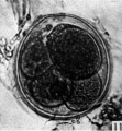
Fig. 11. Photograph of a five-cell stage ovum of the pony (P 4) in agar. x 480.


Plate 3

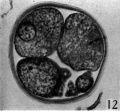
Fig. 12. Section through the five-cell ovum shown in fig. 11. x 520.
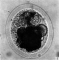
Fig. 13. Photograph of the living morula. of the pony (P 6). it shows extensive deutoplasmolysis. x 480.
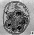
Figs. 14, 15. Two consecutive sections through the morula. shown in fig. 13. x 520.
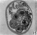
Figs. 14, 15. Two consecutive sections through the morula. shown in fig. 13. x 520.


Genital Development
Gastrointestinal Tract
Prenatal Development of the Digestive System in the Horse.[3]
- 21 days - oral cavity was an empty space, and the liver contained proliferating endodermal cells.
- 25 days - fusiform stomach and the pancreatic bud were present.
- 28 days - small tongue and the esophagus occurred.
- 30 days - primary and secondary palates were developed, the liver contained cords of hepatocytes, and the pancreas was triangular.
- 40 days - crypts had formed in the intestinal loops, cell differentiation was observed in the hepatic parenchyma, and the pancreas was elongated.
- 50 days - Pancreatic acini and islets and intestines were highly convoluted.
- 75 days - Three segments of the pharynx
- 105 days - intestinal villi were wide with round tips; especially, the liver, stomach, and oral cavity showed key steps of anatomical and cellular differentiation.
References
- ↑ <pubmed>22645258</pubmed>
- ↑ Hamilton WJ. Cleavage Stages of the Ova of the Horse, with Notes on Ovulation. J Anat. 1945 Jul; 79(Pt 3): 127–130.3.
- ↑ <pubmed>24778084</pubmed>
Reviews
<pubmed></pubmed> <pubmed></pubmed> <pubmed></pubmed> <pubmed></pubmed>
Articles
<pubmed></pubmed> <pubmed>24778084</pubmed> <pubmed>22645258</pubmed> <pubmed>21923925</pubmed> <pubmed>21209420</pubmed>
Search Pubmed
Search Pubmed: equine development
External Links
External Links Notice - The dynamic nature of the internet may mean that some of these listed links may no longer function. If the link no longer works search the web with the link text or name. Links to any external commercial sites are provided for information purposes only and should never be considered an endorsement. UNSW Embryology is provided as an educational resource with no clinical information or commercial affiliation.
- Oklahoma State University Learning Reproduction in Farm Animals
| Animal Development: axolotl | bat | cat | chicken | cow | dog | dolphin | echidna | fly | frog | goat | grasshopper | guinea pig | hamster | horse | kangaroo | koala | lizard | medaka | mouse | opossum | pig | platypus | rabbit | rat | salamander | sea squirt | sea urchin | sheep | worm | zebrafish | life cycles | development timetable | development models | K12 |
Glossary Links
- Glossary: A | B | C | D | E | F | G | H | I | J | K | L | M | N | O | P | Q | R | S | T | U | V | W | X | Y | Z | Numbers | Symbols | Term Link
Cite this page: Hill, M.A. (2024, April 19) Embryology Horse Development. Retrieved from https://embryology.med.unsw.edu.au/embryology/index.php/Horse_Development
- © Dr Mark Hill 2024, UNSW Embryology ISBN: 978 0 7334 2609 4 - UNSW CRICOS Provider Code No. 00098G