Histology Stains: Difference between revisions
From Embryology
No edit summary |
|||
| Line 198: | Line 198: | ||
===Hematoxylin and Eosin=== | ===Hematoxylin and Eosin=== | ||
Acronym "H and E" stain. | |||
<gallery> | <gallery> | ||
Revision as of 11:49, 3 April 2010
Introduction
This page gives a general overview of some histological stains used to identify structures in cells and tissues. This stains information should also be considered in relation to fixatives.
Medicine Foundations students do not need to know this detail.
Common Stains and Their Reactions | |||||
| Haematoxylin | mucins - light blue | ||||
| Eosin | colloid - pinkmuscle - red | ||||
| Iron Haematoxylin | |||||
| Van Gieson | muscle: yellow/browncartilage - pink | ||||
| Verhoeff's Elastin | elastic fibres - black | ||||
| Tartrazine | |||||
| Silver Impregnation | reticular fibres - black | ||||
| Methyl Green | |||||
| Nuclear Fast Red | |||||
| Gomori's Trichrome | keratin - redmuscle - purple/red | ||||
| Heidenhain's Azan | muscle - red | ||||
| Osmium tetroxide | myelin, lipids - black | ||||
| Alcian Blue | mucins, - blue | ||||
| Periodic acid-Schiff (PAS) | mucins, glycogen, glycocalyx - magenta | ||||
| PTAH | muscle bands - blue | ||||
| Masson's Trichrome | cartilage, mucins - blue or green; muscle - red | ||||
| Luxol Fast Blue | myelin - blue~ | ||||
| Aldehyde Fuchsin | elastic fibres, mast cells - deep purple | ||||
| Light Green | |||||
| Gallocyanin | nucleic acids, Nissl granules - dark blue | ||||
| Romanowsky(e.g. Leishman's stain) | acidophils - redbasophils - blueazurophilic - purple | ||||
| Aldehyde Pararosanilin | elastic fibres - purple | ||||
Supporting Stain Information
Hematoxylin and Eosin
Acronym "H and E" stain.
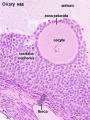
Ovary Histology

Ovary Histology
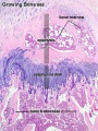
Endochondral ossification


Hematoxylin
- Stains nuclei blue to dark-blue.
- Stains the matrix of hyaline cartilage, myxomatous, and mucoid material pale blue.
- Stains myelin weakly but is not noticeable if combined with eosin stain.
Eosin
- Stains cytoplasm pink to red; red blood cells are also bright red.
- Common counterstain to hematoxylin.
- Stain intensity varies with the formula as well as the fixative.
Periodic acid-Schiff (PAS)
- Stains glycogen, mucin, fungus, basement membrane and other substances.
- Stain used to detect fungal organisms and cytoplasmic accumulation of glycogen.
- Stains lysosomes granules red-purple, can be used in recognition of macrophages.
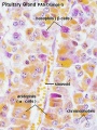
Pituitary histology
Pituitary histology


Alcian Blue
- Stains mucopolysaccharides or glycosaminoglycans
- cationic dye (positively charged molecule) for the demonstration of glycosaminoglycans.
- binds anionic (negative) sites on the polysaccharide.
- can be combined with H&E and VG staining methods.
Masson’s Trichrome Stain
- Stains nuclei deep blue, skeletal and smooth muscles red, collagen and mucin blue.
- Stains brain and spinal cord parenchymal tissue dusky pink to red.
- Used to evaluate fibrosis
- Striations in skeletal muscles also shows up much better in Masson’s trichrome than in hematoxylin and eosin stain.
- Although called a trichrome, four dyes (hematoxylin, Biebrich scarlet, acid fuchsin, and analine blue) are utilized.
PhosphoTungstic Acid Hematoxylin (PTAH)
- Stains nucleus and cytoplasm detail and connective tissue fibers.
- Stains collagen pink, fibrin blue, and striated muscle blue.
- Historic stain used to show CNS reactive astrocytes now used immunochemistry for glial fibrillary acidic protein (GFAP).
Toulidine Blue
- Stains nucleus blue and cytoplasm light blue.
- A synthetic dye in the thiazins family.
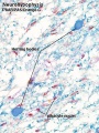
Verhoeff-Van Gieson
- Verhoeff-Van Gieson or elastic-Van Gieson (EVG) stain.
- This is a combination of Verhoeff’s elastic stain which is a hematoxylin stain containing ferric chloride and Wright’s iodine solution and Van Gieson stain which contains acid fuchsin, picric acid, and hematoxylin.
- Stains elastic fibers blue-black to black, collagen pale red, other tissue elements yellow, and nuclei blue to black.
Some text modified from: Theory and practice of histological techniques. 3rd edt. By Bancroft JD and Stevens A. Churchill Livingstone, 1990.