Head Development - Abnormalities
Introduction
Many head and neck structures are derived from pharyngeal arches 1 and 2 which undergo extensive remodelling during head development. Within the head are embedded many other complex developing structures, it is therefore not uncommon for this body region to have many associated abnormalities. Note that for neural and other head organs, look at the relevant section of notes which have their own abnormalities page.
Head and Neck Abnormalities
- Congenital Auricular Sinuses and Cysts
- Pharyngeal Abnormalities - Sinuses, Fistula, Cysts, Vestiges
- Pyriform Sinus Fistula (Piriform Sinus Fistula)
- First Arch Syndrome
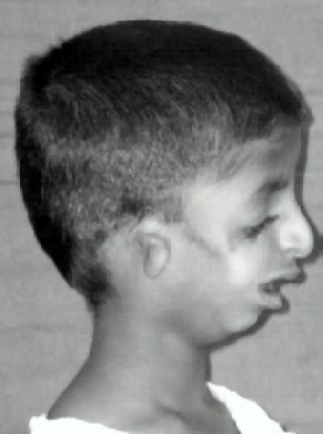
- Treacher Collins syndrome
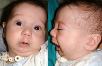
- Pierre Robin syndrome
- DiGeorge syndrome
- Accessory thymic tissue
- Ectopic parathyroid glands
- Thyroid Gland Anomalies
Pharyngeal Abnormalities
There are typically four different terms for the different types of pharyngeal abnormalities, all of these are relatively rare.
Sinuses
A pharyngeal groove defect, when a portion of the groove persists and opens to the skin surface, located laterally on the neck.
Fistula
A pharyngeal membrane defect, a tract extends from pharynx (tonsillar fossa) beween the carotid arteries (internal and external) to open on side of neck.
Cysts
A cervical sinus defect, remants of the cervical sinus remains as a fluid-filled cyst lined by an epithelium.
Vestiges
Cartilaginous or bony developmental remnants that lie under the skin on side of neck.
Clefting
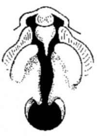
The way in which the upper jaw forms from fusion of the smaller upper prominence of the first pharyngeal arch leads to a common congenital defect in this region called "clefting", which may involve either the upper lip, the palate or both structures.
Cleft Lip
|
|
Cleft Palate
|
(Data: Congenital Malformations Australia 1981-1992 P. Lancaster and E. Pedisich ISSN 1321-8352) Links: Development Animation - Palate 1 | Development Animation - Palate 2 | Orofacial Cleft with or without cleft palate Search Pubmed Now: cleft lip | cleft palate |
First Arch Syndrome
There are 2 major types of genetic first arch syndromes, Treacher Collins and Pierre Robin, both result in extensive facial abnormalites.
Search Pubmed Now: facial cleft
Treacher Collins syndrome (TCS)
- a rare autosomal dominant craniofacial disorder (1:50,000)
- TCOF1 gene encoding Treacle protein
- caused by frameshift deletions or duplications
- located chromosome 5
- encodes a serine/alanine-rich nucleolar phospho-protein
Features
- hypoplasia of the mandible and zygomatic complex
- down-slanting palpebral fissures
- coloboma of the lower eyelid
- absence of eyelashes medial to the defect
- external and middle ear malformation
- conductive hearing loss
Zebrafish Model
Fishing the molecular bases of Treacher Collins syndrome[1]
- "Treacher Collins syndrome (TCS) is an autosomal dominant disorder of craniofacial development, and mutations in the TCOF1 gene are responsible for over 90% of TCS cases. The knowledge about the molecular mechanisms responsible for this syndrome is relatively scant, probably due to the difficulty of reproducing the pathology in experimental animals. Zebrafish is an emerging model for human disease studies, and we therefore assessed it as a model for studying TCS. We identified in silico the putative zebrafish TCOF1 ortholog and cloned the corresponding cDNA. The derived polypeptide shares the main structural domains found in mammals and amphibians. Tcof1 expression is restricted to the anterior-most regions of zebrafish developing embryos, similar to what happens in mouse embryos. Tcof1 loss-of-function resulted in fish showing phenotypes similar to those observed in TCS patients, and enabled a further characterization of the mechanisms underlying craniofacial malformation. Besides, we initiated the identification of potential molecular targets of treacle in zebrafish. We found that Tcof1 loss-of-function led to a decrease in the expression of cellular proliferation and craniofacial development. Together, results presented here strongly suggest that it is possible to achieve fish with TCS-like phenotype by knocking down the expression of the TCOF1 ortholog in zebrafish. This experimental condition may facilitate the study of the disease etiology during embryonic development."
Links: OMIM - Treacher Collins Syndrome | Medline Plus - Treacher Collins Syndrome | PMID 20301704
Pierre Robin syndrome
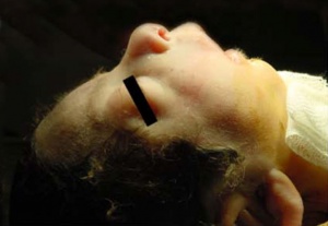
Also called Pierre Robin sequence.
- micrognathia
- retroglossia
- U-shaped posterior cleft palate
Frontal and lateral views of an infant with Pierre Robin sequence.[3]
Links: OMIM - Pierre Robin Syndrome | Medline Plus - Pierre Robin Syndrome | Pierre Robin Network
Mandibular Hypoplasia
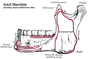
One of the most common malformations of the facial skeleton usually associated with a deficient gonial angle, ascending ramus, and mandibular corpus.
- gonial angle - (angle of the jaw, angle of the mandible) the angle formed by the junction of the posterior and lower borders of the human lower jaw.
- ascending ramus - the more or less vertical part of the jaw which carries the joint with the skull.
- mandibular corpus - the horizontal or tooth-bearing portion of the mandible.
- Links: Growth of the Mandible
Choanal Atresia
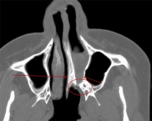
- Choanal atresia is the most common form of congenital nasal obstruction, usually diagnosed at birth.[4]
- failure of the posterior nasal cavity (choanae) to communicate with the nasopharynx.
- Thought to be secondary to an abnormality during the rupture of the buccopharyngeal membrane in the embryological period.
- Links: Smell Development
Cephalic Disorders
Cephalic (Greek, kephale = head) are a group of abnormalities that relate to a wide range of skeletal (skull) and neural (brain) associated defects.
Neural Associated
Anencephaly, Hydrocephalus, Encephalocele, Colpocephaly (occipital horn enlargement), Lissencephaly (smooth brain), Porencephaly (cyst or cavity in cerebral hemisphere), Acephaly (absence of head), Exencephaly (brain outside skull), Macrocephaly (large head), Micrencephaly (small brain),
Skeletal Associated
- Otocephaly - absence of lower jaw.
- Brachycephaly - premature fusion of coronal suture.
- Oxycephaly - premature fusion of coronal suture + other.
- Plagiocephaly - premature unilateral fusion of coronal or lambdoid sutures.
- Scaphocephaly - premature fusion of sagittal suture.
- Trigonocephaly - premature fusion of the metopic suture.
- Links: Skull Development
Fetal Alcohol Syndrome
(FAS) Due to alcohol in early development (week 3+) leading to both facial and neurological abnormalities. This disorder was clinically described (USA) in humans about 30 years ago (1973), while historically alcohol's teratogenic effects were identified in the early 20th century in a mix with the prohibition cause of the period. Similar effects without the obvious alterations to appearance, but with nervous system effects, are sometimes identified as Fetal Alcohol Effects (FAE). Alcohol is able to cross the placenta from maternal circulation through the placenta into fetal circulation.
FAS Features:
- lowered ears, small face, mild+ retardation
- Microcephaly - leads to small head circumference
- Short Palpebral fissure - opening of eye
- Epicanthal folds - fold of skin at inside of corner of eye
- Flat midface
- Low nasal bridge
- Indistinct Philtrum - vertical grooves between nose and mouth
- Thin upper lip
- Micrognathia - small jaw
Exposure of embryos in vitro to ethanol simulates premature differentiation of prechondrogenic mesenchyme of the facial primordia (1999)
References
- ↑ <pubmed>22295061</pubmed>
- ↑ <pubmed>20716376</pubmed>| PMC2931455 | BMC Pregnancy Childbirth.
- ↑ <pubmed>22300418</pubmed>| Ital J Pediatr.
- ↑ 4.0 4.1 <pubmed>21772853</pubmed>
Reviews
<pubmed></pubmed>
Articles
Search Pubmed
Search Pubmed: abnormal head development | abnormal skull development | abnormal skull development
Glossary Links
- Glossary: A | B | C | D | E | F | G | H | I | J | K | L | M | N | O | P | Q | R | S | T | U | V | W | X | Y | Z | Numbers | Symbols | Term Link
Cite this page: Hill, M.A. (2024, April 16) Embryology Head Development - Abnormalities. Retrieved from https://embryology.med.unsw.edu.au/embryology/index.php/Head_Development_-_Abnormalities
- © Dr Mark Hill 2024, UNSW Embryology ISBN: 978 0 7334 2609 4 - UNSW CRICOS Provider Code No. 00098G
