Granulosa cell: Difference between revisions
mNo edit summary |
mNo edit summary |
||
| (6 intermediate revisions by the same user not shown) | |||
| Line 3: | Line 3: | ||
[[File:Ovary histology 061.jpg|thumb|300px|alt=Oocyte and developing zona pellucida in the ovary|Oocyte and developing granolas cell layer]] | [[File:Ovary histology 061.jpg|thumb|300px|alt=Oocyte and developing zona pellucida in the ovary|Oocyte and developing granolas cell layer]] | ||
Surrounding the oocyte as it develops within the ovary follicle are multiple layers of granulosa | Surrounding the oocyte as it develops within the ovary follicle are multiple layers of {{granulosa cell}}s that are bound to the thick specialised extracellular matrix, the {{zona pellucida}}. The innermost layer of these cells, the corona radiata, communicate directly with the oocyte by cytoplasmic extensions passing through the {{zona pellucida}}. Following release of the oocyte at ovulation, these cells form the granolas layer. | ||
Granulosa cells can also have specific names depending upon location within the ovarian follicle: cumulus oophrous (Latin, ''cumulus'' = a little mound; Greek, ''oo''= egg, ''phorus'' = carrying) also called cumulus granulosa cells directly around the zone pellucida and released with the oocyte; '''membrana granulosa''' also called mural granulosa cells forming the layer within the follicle antral wall; '''discus proligerus''' can refer to the attachment between cumulus oophrous and membrane granulosa; and '''mural granulosa cells''' that line the follicular wall. | Granulosa cells can also have specific names depending upon location within the ovarian follicle: cumulus oophrous (Latin, ''cumulus'' = a little mound; Greek, ''oo''= egg, ''phorus'' = carrying) also called cumulus granulosa cells directly around the zone pellucida and released with the oocyte; "corona radiata" forming the initial layer in contact with the zone pellucida; '''membrana granulosa''' also called mural granulosa cells forming the layer within the follicle antral wall; '''discus proligerus''' can refer to the attachment between cumulus oophrous and membrane granulosa; and '''mural granulosa cells''' that line the follicular wall. | ||
| Line 14: | Line 14: | ||
{{Fertilization Links}} | {{Fertilization Links}} | ||
==Some Recent Findings== | ==Some Recent Findings== | ||
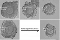
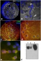
[[File:Mouse_germinal_vesicle_04.jpg|thumb|alt=Mouse germinal vesicle|Mouse germinal vesicle with granulosa layer | [[File:Mouse_germinal_vesicle_04.jpg|thumb|alt=Mouse germinal vesicle|Mouse germinal vesicle with granulosa layer{{#pmid:25144310|PMID25144310}}]] | ||
{| | {| | ||
|-bgcolor="F5FAFF" | |-bgcolor="F5FAFF" | ||
| | | | ||
* '''Transcription profile of the insulin-like growth factor signaling pathway during human ovarian follicular development'''{{#pmid:30877600|PMID30877600}} "The IGF signaling cascade exerts important regulatory functions in human ovarian folliculogenesis. The scope of this study was to evaluate the transcription profile of insulin-like growth factor (IGF) genes during human ovarian follicle development and to analyze follicle fluid levels of key IGF proteins. Gene expression profiling was performed with microarray gene analysis. The analysis was assessed from ovarian follicles and granulosa cells (GCs) obtained from isolated stage-specific human ovarian follicles, including preantral follicles, small antral follicles, and preovulatory follicles. Numerous genes involved in the IGF signaling pathway was evaluated and key genes were validated by qPCR from GCs. Protein levels of various IGF components of human follicular fluid (FF) were measured by ELISA and time-resolved immunofluorometric assays (TRIFMA). The gene expression levels of PAPPA, IGF2, IGF receptors and intracellular IGF-activated genes increased with increasing follicle size. This was especially prominent in the late preovulatory stage where IGF2 expression peaked. Protein levels of intact IGF binding protein-4 decreased significantly in FF from large preovulatory follicles compared with small antral follicles concomitant with higher protein levels of PAPP-A. The IGF modulators IGF-2 receptor, IGFBPs, stanniocalcins, and IGF-2 mRNA binding proteins were all observed to be expressed in the different follicle stages. This study confirms and highlights the importance of PAPP-A regulating bioactive IGF levels throughout folliculogenesis and especially for the high rate of granulosa cell proliferation and expression of key ovarian hormones important in the last part of the follicular phase of the menstrual cycle." | |||
|} | |} | ||
{| class="wikitable mw-collapsible mw-collapsed" | {| class="wikitable mw-collapsible mw-collapsed" | ||
! More recent papers | ! More recent papers | ||
|- | |- | ||
| [[File:Mark_Hill.jpg|90px|left]] {{Most_Recent_Refs}} | | [[File:Mark_Hill.jpg|90px|left]] {{Most_Recent_Refs}} | ||
| Line 30: | Line 29: | ||
Search term: [http://www.ncbi.nlm.nih.gov/pubmed/?term=Granulosa+cell+development ''Granulosa cell development''] | Search term: [http://www.ncbi.nlm.nih.gov/pubmed/?term=Granulosa+cell+development ''Granulosa cell development''] | ||
|} | |||
{| class="wikitable mw-collapsible mw-collapsed" | |||
! Older papers | |||
|- | |||
| {{Older papers}} | |||
* '''Transcriptomic Diversification of Developing Cumulus and Mural Granulosa Cells in Mouse Ovarian Follicles'''{{#pmid:25376232|PMID25376232}} "Cumulus cells and mural granulosa cells (MGCs) have functionally distinct roles in antral follicles, and comparison of their transcriptomes at a global and systems level can propel future studies on mechanisms underlying their functional diversity. ...Gene Ontology analysis revealed that cumulus cells of small follicles were enriched in transcripts generally associated with catalytic components of metabolic processes, while those from large follicles were involved in regulation of metabolism, cell differentiation, and adhesion. Contrast of cumulus cells versus MGCs revealed that cumulus cells were enriched in transcripts associated with metabolism and cell proliferation while MGCs were enriched for transcripts involved in cell signaling and differentiation. In-vitro and in-vivo models were used to test the hypothesis that higher levels of transcripts in cumulus cells versus MGCs is the result of stimulation by oocyte-derived paracrine factors (ODPFs)." | |||
* '''Wt1 functions in ovarian follicle development by regulating granulosa cell differentiation'''{{#pmid:24009315|PMID24009315}} "The Wt1 gene encodes a nuclear transcription factor that is specifically expressed in ovarian granulosa cells. However, the physiological significance of Wt1 in ovarian follicle development remains elusive. ...The results of in vitro studies demonstrated that the expression of follicle stimulating hormone receptor (FSHR), 3β-hydroxysteroid dehydrogenase and Aromatase was inhibited by Wt1 in granulosa cells, and mutation of Wt1 resulted in the upregulation of these genes and in the premature differentiation of granulosa cells. We also found that Wt1 was likely involved in granulosa cell development via the regulation of E-cadherin and Par6b expression. Mutation in Wt1 caused defects in polarity establishment in granulosa cells, which also likely contributed to the observed aberrant follicle development." | |||
|} | |} | ||
==Follicle Granulosa Cells== | ==Follicle Granulosa Cells== | ||
| Line 46: | Line 52: | ||
Cumulus granulosa cells lie directly around the oocyte zone pellucida and are released with the oocyte at ovulation. These cells have been shown to exchange biological signals with oocytes through the gap junctions. | Cumulus granulosa cells lie directly around the oocyte zone pellucida and are released with the oocyte at ovulation. These cells have been shown to exchange biological signals with oocytes through the gap junctions. | ||
[[File:Oocyte and cumulus granulosa signaling 01.jpg|600px]] | |||
Oocyte and Cumulus Granulosa Signaling{{#pmid:23382188|PMID23382188}} | |||
| Line 56: | Line 67: | ||
Mural Granulosa Cells (MGCs) lining the ovulating follicle remain within the ovary and differentiate to form part of the corpus luteum. | Mural Granulosa Cells (MGCs) lining the ovulating follicle remain within the ovary and differentiate to form part of the corpus luteum. | ||
Steroid hormone synthesis by mural granulosa cells is dependent upon the multiligand receptor LOX-1. | Steroid hormone synthesis by mural granulosa cells is dependent upon the multiligand receptor LOX-1.{{#pmid:24710763|PMID24710763}} | ||
{| | {| | ||
| Line 73: | Line 84: | ||
:'''Links:''' [[Ovary_Development#Corpus_Luteum|Corpus luteum]] | :'''Links:''' [[Ovary_Development#Corpus_Luteum|Corpus luteum]] | ||
==Follicle Growth== | |||
[[File:Ovary follicle size graph.jpg|alt=Ovary follicle size graph|600px]] | |||
Graph shows species comparison in follicle size growth (diameter) at different stages of follicle development.{{#pmid:16509981|PMID16509981}} (See also [[:File:Ovary oocyte size graph.jpg|Oocyte size graph]]) | |||
{{Follicle class collapse table}} | |||
==Mouse Granulosa Cells== | ==Mouse Granulosa Cells== | ||
| Line 82: | Line 100: | ||
==Molecular== | ==Molecular== | ||
[[File:Sheep follicle gene expression 01.jpg|600px]] | |||
{| | |||
| Granulosa gene expression (sheep){{#pmid:26540452|PMID26540452}} | |||
| width=300px| | |||
* <font color=red>'''red'''</font> - genes expressed in granulose cells. | |||
* <font color=blue>'''blue'''</font> - genes expressed in oocytes. | |||
* '''black''' - genes expressed both in oocytes and granulosa cells. | |||
|} | |||
{| | {| | ||
| | | | ||
| Line 90: | Line 118: | ||
'''Signaling Factors''' | '''Signaling Factors''' | ||
* FOXL2 - (forkhead transcription factor) expressed in the less differentiated granulosa cells of small and medium follicles. | * FOXL2 - (forkhead transcription factor) expressed in the less differentiated granulosa cells of small and medium follicles.{{#pmid:25369636|PMID25369636}} [http://omim.org/entry/605597 OMIM605597] | ||
* HAS2, PTGS2, PTX3, TNFAIP6 | * HAS2, PTGS2, PTX3, TNFAIP6 | ||
* | * {{BMP}}15, GDF9, {{FGF}}8 | ||
'''Kisspeptin''' | |||
In the {{pig}} ovary kisspeptin and its receptor are expressed by follicle cells.{{#pmid:29698886|PMID29698886}} Kisspeptin, acting through its receptor KISS1R, have a key neuroendocrine role in the regulation of mammalian reproduction. It is not known what specific effect this factor may have on follicle development. | |||
{{Factor Links}} | {{Factor Links}} | ||
| [[File:Ovarian follicle molecular interactions 01.jpg|thumb|300px|alt=Ovarian follicle molecular interactions|Molecular paracrine interactions | | [[File:Ovarian follicle molecular interactions 01.jpg|thumb|300px|alt=Ovarian follicle molecular interactions|Molecular paracrine interactions{{#pmid:24711992|PMID24711992}}]] | ||
|} | |} | ||
==Abnormalities== | ==Abnormalities== | ||
===Granulosa cell tumour=== | ===Granulosa cell tumour=== | ||
| Line 107: | Line 140: | ||
[[File:Granulosa cell tumour 01.jpg|300px]] | [[File:Granulosa cell tumour 01.jpg|300px]] | ||
Photograph of a granulosa cell tumour | Photograph of a granulosa cell tumour{{#pmid:21060767|PMID21060767}} | ||
| Line 116: | Line 149: | ||
===Reviews=== | ===Reviews=== | ||
{{#pmid:15271471}} | |||
{{#pmid:14967918}} | |||
===Articles=== | ===Articles=== | ||
{{#pmid:24422759}} | |||
{{#pmid:24270888}} | |||
{{#pmid:12077402}} | |||
===Search Pubmed=== | ===Search Pubmed=== | ||
| Line 138: | Line 170: | ||
==Additional Images== | ==Additional Images== | ||
<gallery> | <gallery> | ||
File:Oocyte and cumulus granulosa signaling 01.jpg|Oocyte and Cumulus Granulosa Signaling | |||
File:Oocyte and cumulus granulosa signaling 02.jpg|Oocyte and Cumulus Granulosa Signaling Human and Sheep | |||
File:Oocyte and cumulus granulosa signaling 03.jpg|Oocyte and Cumulus Granulosa Signaling Mouse and Rat | |||
File:Cat_spermatozoa_bound_to_oocyte_zona_pellucida.jpg|[[Cat Development|Cat]] oocyte zona pellucida spermatozoa bound SEM | File:Cat_spermatozoa_bound_to_oocyte_zona_pellucida.jpg|[[Cat Development|Cat]] oocyte zona pellucida spermatozoa bound SEM | ||
File:Hamster_oocyte_zona_pellucida_SEM.jpg|[[Hamster_Development|Hamster]] oocyte zona pellucida SEM | File:Hamster_oocyte_zona_pellucida_SEM.jpg|[[Hamster_Development|Hamster]] oocyte zona pellucida SEM | ||
| Line 146: | Line 181: | ||
==Terms== | ==Terms== | ||
* '''acrosome reaction''' - The chemical change within the [[S#spermatozoa|spermatozoa]] following binding to the zona pellucida, that leads to the release of acrosomal enzymatic contents. These enzymes degrade the zona pellucida and allow a [[S#spermatozoa|spermatozoa]] to penetrate an oocyte. | * '''acrosome reaction''' - The chemical change within the [[S#spermatozoa|spermatozoa]] following binding to the zona pellucida, that leads to the release of acrosomal enzymatic contents. These enzymes degrade the zona pellucida and allow a [[S#spermatozoa|spermatozoa]] to penetrate an oocyte. | ||
* '''Call–Exner bodies''' - small histologically [[Histology_Stains#Eosin|eosinophilic]] fluid-filled spaces between granulosa cells. Named after Emma Louise Call (1847 - 1937) one of the first American women physician and Sigmund Exner (1846 – 1926) an Austrian physiologist. | * '''Call–Exner bodies''' - small histologically [[Histology_Stains#Eosin|eosinophilic]] fluid-filled spaces between granulosa cells. Named after Emma Louise Call (1847 - 1937) one of the first American women physician and Sigmund Exner (1846 – 1926) an Austrian physiologist. | ||
* '''cumulus cell''' - (Latin, ''cumulus'' = a little mound; Greek, ''oo''= egg, phorus=carrying) granulosa cells directly around the zone pellucida | |||
* '''cumulus cell''' - (Latin, ''cumulus'' = a little mound; Greek, ''oo''= egg, phorus=carrying) granulosa cells directly around the zone pellucida. | |||
* '''granulosa cell''' - A specific cell type that proliferates in association with the oocyte within the developing [[F#follicle|follicles]] of the [[O#ovary|ovary]]. These cells form the [[F#follicle|follicle]] stratum granulosa and are also given specific names based upon their position within the [[F#follicle|follicle]]. In the [[A#antral_follicle|antral follicle]], [[M#membrana_granulosa|membrana granulosa]] sits on the [[F#follicular_basal_lamina|follicular basal lamina]] and lines the antrum as a stratified epithelium. The [[C#cumulus_oophorus|cumulus oophorus]] is a column of granulosa cells that attaches the oocyte to the [[F#follicle|follicle]] wall. The [[C#corona_radiata|corona radiata]] are the [[G#granulosa cell|granulosa cells]] that directly surround the oocyte, and are released along with it at ovulation. Following ovulation the corona radiata provide physical protection to the oocyte and granulosa cells within the ovulating [[F#follicle|follicle]] contribute to corpus luteum. | * '''granulosa cell''' - A specific cell type that proliferates in association with the oocyte within the developing [[F#follicle|follicles]] of the [[O#ovary|ovary]]. These cells form the [[F#follicle|follicle]] stratum granulosa and are also given specific names based upon their position within the [[F#follicle|follicle]]. In the [[A#antral_follicle|antral follicle]], [[M#membrana_granulosa|membrana granulosa]] sits on the [[F#follicular_basal_lamina|follicular basal lamina]] and lines the antrum as a stratified epithelium. The [[C#cumulus_oophorus|cumulus oophorus]] is a column of granulosa cells that attaches the oocyte to the [[F#follicle|follicle]] wall. The [[C#corona_radiata|corona radiata]] are the [[G#granulosa cell|granulosa cells]] that directly surround the oocyte, and are released along with it at ovulation. Following ovulation the corona radiata provide physical protection to the oocyte and granulosa cells within the ovulating [[F#follicle|follicle]] contribute to corpus luteum. | ||
* '''mural granulosa cell''' - (MGC) granulosa cells that line the follicular wall and have an endocrine function. | * '''mural granulosa cell''' - (MGC) granulosa cells that line the follicular wall and have an endocrine function. | ||
Latest revision as of 14:55, 25 March 2019
| Embryology - 24 Apr 2024 |
|---|
| Google Translate - select your language from the list shown below (this will open a new external page) |
|
العربية | català | 中文 | 中國傳統的 | français | Deutsche | עִברִית | हिंदी | bahasa Indonesia | italiano | 日本語 | 한국어 | မြန်မာ | Pilipino | Polskie | português | ਪੰਜਾਬੀ ਦੇ | Română | русский | Español | Swahili | Svensk | ไทย | Türkçe | اردو | ייִדיש | Tiếng Việt These external translations are automated and may not be accurate. (More? About Translations) |
Introduction
Surrounding the oocyte as it develops within the ovary follicle are multiple layers of granulosa cells that are bound to the thick specialised extracellular matrix, the zona pellucida. The innermost layer of these cells, the corona radiata, communicate directly with the oocyte by cytoplasmic extensions passing through the zona pellucida. Following release of the oocyte at ovulation, these cells form the granolas layer.
Granulosa cells can also have specific names depending upon location within the ovarian follicle: cumulus oophrous (Latin, cumulus = a little mound; Greek, oo= egg, phorus = carrying) also called cumulus granulosa cells directly around the zone pellucida and released with the oocyte; "corona radiata" forming the initial layer in contact with the zone pellucida; membrana granulosa also called mural granulosa cells forming the layer within the follicle antral wall; discus proligerus can refer to the attachment between cumulus oophrous and membrane granulosa; and mural granulosa cells that line the follicular wall.
In human development, during the first week of development following fertilization the granolsa cell layer and zona pellucida remain surrounding the blastocyst from which it "hatches" to commence implantation.
Some Recent Findings

|
| More recent papers |
|---|
 This table allows an automated computer search of the external PubMed database using the listed "Search term" text link.
More? References | Discussion Page | Journal Searches | 2019 References | 2020 References Search term: Granulosa cell development |
| Older papers |
|---|
| These papers originally appeared in the Some Recent Findings table, but as that list grew in length have now been shuffled down to this collapsible table.
See also the Discussion Page for other references listed by year and References on this current page.
|
Follicle Granulosa Cells
Granulosa cells can also have specific names depending upon location within the follicle.
|

|
Cumulus Granulosa Cells

cumulus oophrous (Latin, cumulus = a little mound; Greek, oo = egg, phorus = carrying)
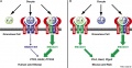
Cumulus granulosa cells lie directly around the oocyte zone pellucida and are released with the oocyte at ovulation. These cells have been shown to exchange biological signals with oocytes through the gap junctions.

Oocyte and Cumulus Granulosa Signaling[5]
Search PubMed term: Cumulus Granulosa development
<pubmed limit=5>Cumulus Granulosa development</pubmed>
Mural Granulosa Cells
Mural Granulosa Cells (MGCs) lining the ovulating follicle remain within the ovary and differentiate to form part of the corpus luteum.
Steroid hormone synthesis by mural granulosa cells is dependent upon the multiligand receptor LOX-1.[6]
| Mural Granulosa Cells | Corpus Luteum Granulosa cells |
|---|---|

|

|
Search PubMed term: Mural Granulosa development
<pubmed limit=5>Mural Granulosa development</pubmed>
- Links: Corpus luteum
Follicle Growth

Graph shows species comparison in follicle size growth (diameter) at different stages of follicle development.[7] (See also Oocyte size graph)
| Human Ovarian Follicle Classification | |||||
| Class | Alternate nomenclature | Type | Number of Cells | Size (diameter µm) | Size ultrasound (mm) |
|---|---|---|---|---|---|
| primordial follicle | small | 1, 2, 3 | 25 | less than 50 | |
| primary follicle | preantral | 4 5 |
26 - 100 101 - 300 |
up to 200 | |
| secondary follicle | antral small antral large antral |
6 7 |
3001 - 500 501 - 1000 |
500 1000 - 6000 |
less than 18 |
| preovulatory follicle | Graafian | 8 | greater than 1000 | greater than 6000 | 18 – 28 |
| Links: ovary | oocyte | menstrual cycle | |||||
Mouse Granulosa Cells
Oocyte and granulosa cells
Oocyte and granulosa cells
Oocyte and granulosa cells


Molecular

| Granulosa gene expression (sheep)[8] |
|
|
Granulosa Cell Markers
In the pig ovary kisspeptin and its receptor are expressed by follicle cells.[10] Kisspeptin, acting through its receptor KISS1R, have a key neuroendocrine role in the regulation of mammalian reproduction. It is not known what specific effect this factor may have on follicle development.
|
 Molecular paracrine interactions[11] |
Abnormalities
Granulosa cell tumour
An uncommon non-epithelial cancer of the ovary.

Photograph of a granulosa cell tumour[12]
References
- ↑ Zhou HX, Ma YZ, Liu YL, Chen Y, Zhou CJ, Wu SN, Shen JP & Liang CG. (2014). Assessment of mouse germinal vesicle stage oocyte quality by evaluating the cumulus layer, zona pellucida, and perivitelline space. PLoS ONE , 9, e105812. PMID: 25144310 DOI.
- ↑ Bøtkjær JA, Pors SE, Petersen TS, Kristensen SG, Jeppesen JV, Oxvig C & Andersen CY. (2019). Transcription profile of the insulin-like growth factor signaling pathway during human ovarian follicular development. J. Assist. Reprod. Genet. , , . PMID: 30877600 DOI.
- ↑ Wigglesworth K, Lee KB, Emori C, Sugiura K & Eppig JJ. (2015). Transcriptomic diversification of developing cumulus and mural granulosa cells in mouse ovarian follicles. Biol. Reprod. , 92, 23. PMID: 25376232 DOI.
- ↑ Gao F, Zhang J, Wang X, Yang J, Chen D, Huff V & Liu YX. (2014). Wt1 functions in ovarian follicle development by regulating granulosa cell differentiation. Hum. Mol. Genet. , 23, 333-41. PMID: 24009315 DOI.
- ↑ Peng J, Li Q, Wigglesworth K, Rangarajan A, Kattamuri C, Peterson RT, Eppig JJ, Thompson TB & Matzuk MM. (2013). Growth differentiation factor 9:bone morphogenetic protein 15 heterodimers are potent regulators of ovarian functions. Proc. Natl. Acad. Sci. U.S.A. , 110, E776-85. PMID: 23382188 DOI.
- ↑ Weitzel JM, Vernunft A, Krüger B, Plinski C & Viergutz T. (2014). Inactivation of the LOX-1 pathway promotes the Golgi apparatus during cell differentiation of mural granulosa cells. J. Cell. Physiol. , 229, 1946-51. PMID: 24710763 DOI.
- ↑ Griffin J, Emery BR, Huang I, Peterson CM & Carrell DT. (2006). Comparative analysis of follicle morphology and oocyte diameter in four mammalian species (mouse, hamster, pig, and human). J. Exp. Clin. Assist. Reprod. , 3, 2. PMID: 16509981 DOI.
- ↑ Bonnet A, Servin B, Mulsant P & Mandon-Pepin B. (2015). Spatio-Temporal Gene Expression Profiling during In Vivo Early Ovarian Folliculogenesis: Integrated Transcriptomic Study and Molecular Signature of Early Follicular Growth. PLoS ONE , 10, e0141482. PMID: 26540452 DOI.
- ↑ Georges A, L'Hôte D, Todeschini AL, Auguste A, Legois B, Zider A & Veitia RA. (2014). The transcription factor FOXL2 mobilizes estrogen signaling to maintain the identity of ovarian granulosa cells. Elife , 3, . PMID: 25369636 DOI.
- ↑ Basini G, Grasselli F, Bussolati S, Ciccimarra R, Maranesi M, Bufalari A, Parillo F & Zerani M. (2018). Presence and function of kisspeptin/KISS1R system in swine ovarian follicles. Theriogenology , 115, 1-8. PMID: 29698886 DOI.
- ↑ Chronowska E. (2014). High-throughput analysis of ovarian granulosa cell transcriptome. Biomed Res Int , 2014, 213570. PMID: 24711992 DOI.
- ↑ Abaid LN, Mosquera-Caro M, Kankus RC & Goldstein BH. (2010). Extraordinarily Prolonged Disease Recurrence in a Granulosa Cell Tumor Patient. Case Rep Oncol , 3, 310-314. PMID: 21060767 DOI.
Reviews
Gilchrist RB, Ritter LJ & Armstrong DT. (2004). Oocyte-somatic cell interactions during follicle development in mammals. Anim. Reprod. Sci. , 82-83, 431-46. PMID: 15271471 DOI.
Senbon S, Hirao Y & Miyano T. (2003). Interactions between the oocyte and surrounding somatic cells in follicular development: lessons from in vitro culture. J. Reprod. Dev. , 49, 259-69. PMID: 14967918
Articles
Hatzirodos N, Irving-Rodgers HF, Hummitzsch K, Harland ML, Morris SE & Rodgers RJ. (2014). Transcriptome profiling of granulosa cells of bovine ovarian follicles during growth from small to large antral sizes. BMC Genomics , 15, 24. PMID: 24422759 DOI.
Chen J, Torcia S, Xie F, Lin CJ, Cakmak H, Franciosi F, Horner K, Onodera C, Song JS, Cedars MI, Ramalho-Santos M & Conti M. (2013). Somatic cells regulate maternal mRNA translation and developmental competence of mouse oocytes. Nat. Cell Biol. , 15, 1415-23. PMID: 24270888 DOI.
Matzuk MM, Burns KH, Viveiros MM & Eppig JJ. (2002). Intercellular communication in the mammalian ovary: oocytes carry the conversation. Science , 296, 2178-80. PMID: 12077402 DOI.
Search Pubmed
Search Pubmed: granulosa cell
Search Images: granulosa cell
Search NCBI Bookshelf Granulosa cell
Additional Images
Oocyte and Cumulus Granulosa Signaling
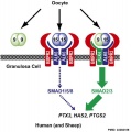
Oocyte and Cumulus Granulosa Signaling Human and Sheep
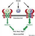
Oocyte and Cumulus Granulosa Signaling Mouse and Rat
Cat oocyte zona pellucida spermatozoa bound SEM
Hamster oocyte zona pellucida SEM
Pig ZPC deposition in oocyte-cumulus complexes
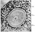
Historic - From a section of the ovary of a 12-year old girl


Terms
- acrosome reaction - The chemical change within the spermatozoa following binding to the zona pellucida, that leads to the release of acrosomal enzymatic contents. These enzymes degrade the zona pellucida and allow a spermatozoa to penetrate an oocyte.
- Call–Exner bodies - small histologically eosinophilic fluid-filled spaces between granulosa cells. Named after Emma Louise Call (1847 - 1937) one of the first American women physician and Sigmund Exner (1846 – 1926) an Austrian physiologist.
- cumulus cell - (Latin, cumulus = a little mound; Greek, oo= egg, phorus=carrying) granulosa cells directly around the zone pellucida.
- granulosa cell - A specific cell type that proliferates in association with the oocyte within the developing follicles of the ovary. These cells form the follicle stratum granulosa and are also given specific names based upon their position within the follicle. In the antral follicle, membrana granulosa sits on the follicular basal lamina and lines the antrum as a stratified epithelium. The cumulus oophorus is a column of granulosa cells that attaches the oocyte to the follicle wall. The corona radiata are the granulosa cells that directly surround the oocyte, and are released along with it at ovulation. Following ovulation the corona radiata provide physical protection to the oocyte and granulosa cells within the ovulating follicle contribute to corpus luteum.
- mural granulosa cell - (MGC) granulosa cells that line the follicular wall and have an endocrine function.
External Links
External Links Notice - The dynamic nature of the internet may mean that some of these listed links may no longer function. If the link no longer works search the web with the link text or name. Links to any external commercial sites are provided for information purposes only and should never be considered an endorsement. UNSW Embryology is provided as an educational resource with no clinical information or commercial affiliation.
- Granulosa Cell tumour Foundation New Zealand
- The Jackson Labs John Eppig
Glossary Links
- Glossary: A | B | C | D | E | F | G | H | I | J | K | L | M | N | O | P | Q | R | S | T | U | V | W | X | Y | Z | Numbers | Symbols | Term Link
Cite this page: Hill, M.A. (2024, April 24) Embryology Granulosa cell. Retrieved from https://embryology.med.unsw.edu.au/embryology/index.php/Granulosa_cell
- © Dr Mark Hill 2024, UNSW Embryology ISBN: 978 0 7334 2609 4 - UNSW CRICOS Provider Code No. 00098G