Gastrointestinal Tract - Liver Development: Difference between revisions
| Line 98: | Line 98: | ||
== Bile Secretion == | == Bile Secretion == | ||
The epithelial cells that line the bile ducts are called '''cholangiocytes'''. | The epithelial cells that line the bile ducts are called '''cholangiocytes'''. | ||
The pathway below describes the production and passage of bile for final excretion into the duodenum: | |||
secreted into '''bile canaliculi''' | # '''hepatocytes''' produce bile | ||
# secreted into '''bile canaliculi''' | |||
connected to '''intrahepatic bile ducts''' | # connected to '''intrahepatic bile ducts''' | ||
# intrahepatic bile ducts connect to the '''hepatic duct''' | |||
intrahepatic bile ducts connect to the '''hepatic duct''' | # then the '''cystic duct''' for storage in the '''gallbladder''' | ||
# then the '''common bile duct''' into the duodenum | |||
then the '''cystic duct''' for storage in the '''gallbladder''' | |||
then the '''common bile duct''' into the duodenum | |||
The term '''extrahepatic bile ducts''' (EHBDs) is used to describe the hepatic, cystic, and common bile ducts. | The term '''extrahepatic bile ducts''' (EHBDs) is used to describe the hepatic, cystic, and common bile ducts. | ||
Revision as of 13:45, 22 August 2010
Introduction
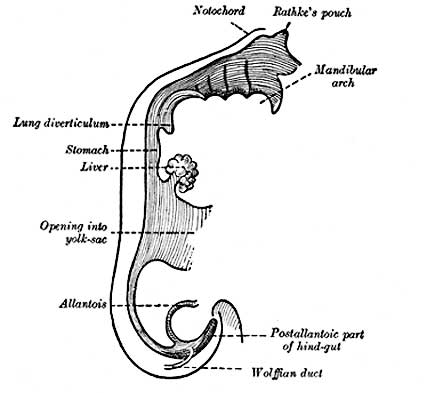
This section of notes gives an overview of how the liver develops. The transverse septum (septum transversum) arises at an embryonic junctional site. The junctional region externally is where the ectoderm of the amnion meets the endoderm of the yolk sac. The junctional region internally is where the foregut meets the midgut. The mesenchymal structure of the transverse septum provides a support within which both blood vessels and the liver begin to form. This structure grows rapidly.
Liver Development Stages
| Feature | |
| hepatic diverticulum development | |
| cell differentiation
septum transversum forming liver stroma hepatic diverticulum forming hepatic trabeculae | |
| epithelial cord proliferation enmeshing stromal capillaries | |
| hepatic gland and its vascular channels enlarge
hematopoietic function appeared | |
| obturation due to epithelial proliferation
bile ducts became reorganized (continuity between liver cells and gut) | |
| biliary ductules developed in periportal connective tissue
produces ductal plates that receive biliary capillaries (More? Timeline human development) |
Data from Godlewski G, etal.[1] "Stage 11 was characterized by hepatic diverticulum development, stage 12 and thereafter by cellular differentiation (septum transversum giving the liver stroma and hepatic diverticulum the hepatic trabeculae), and stage 13 by epithelial cord proliferation enmeshing stromal capillaries. From stage 14, the hepatic gland and its vascular channels presented considerable enlargement while hematopoietic function appeared. From this stage, the development of cystic primordium, never present in rat, was constant in man. At stage 18, after a period of obturation due to epithelial proliferation, the bile ducts became reorganized and ensured the continuity between liver cells and gut. From stages 18 to 23, biliary ductules developed in periportal connective tissue producing ductal plates that received biliary capillaries."
See also Liver development in the rat during the embryonic period (Carnegie stages 15-23).[2]
Components of Liver Formation
Primitive Endoderm
|
|
|
Data from mouse [3]
- Links: Endoderm | Mouse Development
Ductal Plate
The ductal plate is a primitive biliary epithelium which develops in mesenchyme adjacent to portal vein branches (periportal hepatoblasts). During liver development it is extensively reorganised (ductal plate remodelling) within the developing liver to form the intrahepatic bile ducts (IHBD). If remodelling does not occur, leading to excess of embryonic bile duct structures in the portal tract, these developmental abnormalities are described as "ductal plate malformation" (DPM).
Ductal Plate Malformations
- Interlobular bile ducts - autosomal recessive polycystic kidney disease
- Smaller interlobular ducts - von Meyenburg complexes
- Larger intrahepatic bile ducts - Caroli's disease
Bile Secretion
The epithelial cells that line the bile ducts are called cholangiocytes.
The pathway below describes the production and passage of bile for final excretion into the duodenum:
- hepatocytes produce bile
- secreted into bile canaliculi
- connected to intrahepatic bile ducts
- intrahepatic bile ducts connect to the hepatic duct
- then the cystic duct for storage in the gallbladder
- then the common bile duct into the duodenum
The term extrahepatic bile ducts (EHBDs) is used to describe the hepatic, cystic, and common bile ducts.
The developing bile ducts express VEGF while hepatoblasts express angiopoietin-1, these two signals are thought to regulate arterial vasculogenesis and remodeling of the hepatic artery respectively.[4]
References
Reviews
Search Pubmed
Search Bookshelf Liver Development
Search Pubmed Now: Liver Development | Embryonic Liver Development
Glossary Links
- Glossary: A | B | C | D | E | F | G | H | I | J | K | L | M | N | O | P | Q | R | S | T | U | V | W | X | Y | Z | Numbers | Symbols | Term Link
Cite this page: Hill, M.A. (2024, April 19) Embryology Gastrointestinal Tract - Liver Development. Retrieved from https://embryology.med.unsw.edu.au/embryology/index.php/Gastrointestinal_Tract_-_Liver_Development
- © Dr Mark Hill 2024, UNSW Embryology ISBN: 978 0 7334 2609 4 - UNSW CRICOS Provider Code No. 00098G
