Gastrointestinal Tract - Histology: Difference between revisions
No edit summary |
mNo edit summary |
||
| (24 intermediate revisions by the same user not shown) | |||
| Line 1: | Line 1: | ||
{{Header}} | |||
==Introduction== | ==Introduction== | ||
It would be difficult to show all associated histology on a single page as there are many different components of the gastrointestinal tract and associated organs. Therefore this page should be considered as simply a start page to explore more specific topics shown below. | It would be difficult to show all associated histology on a single page as there are many different components of the gastrointestinal tract and associated organs. Therefore this page should be considered as simply a start page containing image galleries and links to explore more specific topics shown below. | ||
Please note that most of these histology images are '''adult tissues''' and may be representative sections from '''other species'''. | |||
Opening a specific image from the gallery will provide additional description notes, species, staining and links to associated resources. | |||
{{Gastrointestinal Tract Links}} | |||
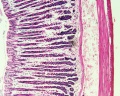
==Oesophagus== | ==Oesophagus== | ||
| Line 14: | Line 19: | ||
File:Oesophagus MALT.jpg | File:Oesophagus MALT.jpg | ||
</gallery> | </gallery> | ||
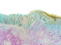
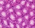
==Stomach== | |||
<gallery> | |||
File:Stomach gastric gland distribution.jpg|Stomach gastric gland distribution cartoon | |||
File:Stomach_histology_001.jpg|stomach labeled overview | |||
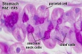
File:Stomach_histology_002.jpg|parietal cells - chief cells | |||
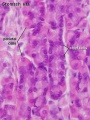
File:Stomach_histology_003.jpg|mucus neck - parietal cells - chief cells | |||
File:Stomach_histology_004.jpg|stomach overview | |||
File:Stomach_histology_005.jpg|stomach mucosa | |||
File:Stomach_histology_006.jpg|mucosa - secretory epithelial sheath - goblet cell | |||
File:Stomach_histology_007.jpg|gastric glands - parietal cells - chief cells | |||
:File:Stomach_histology_008.jpg|stomach overview | |||
</gallery> | |||
{{Stomach Histology}} | |||
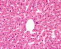
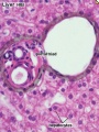
==Liver== | |||
<gallery> | |||
File:Liver_histology_001.jpg|Central vein (label) | |||
File:Liver_histology_101.jpg|Central vein (unlabel) | |||
File:Liver_histology_002.jpg|Portal triad 1 (label) | |||
File:Liver_histology_003.jpg|Portal triad 2 (label) | |||
File:Liver_histology_102.jpg|Portal triad (unlabel) | |||
File:Liver histology 103.jpg|Hepatocytes (unlabel) | |||
File:Liver_histology_004.jpg|Hepatocytes polyploid (label) | |||
</gallery> | |||
{{Liver Histology Images}} | |||
==Gall Bladder== | |||
<gallery> | |||
File:Gall bladder histology 001.jpg|overview (label) | |||
File:Gall bladder histology 003.jpg|overview (unlabel) | |||
File:Gall bladder histology 002.jpg|epithelium (label) | |||
File:Gall bladder histology 004.jpg|epithelium (unlabel) | |||
</gallery> | |||
{{Gall Bladder Histology Images}} | |||
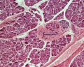
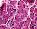
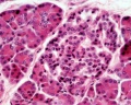
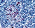
==Pancreas== | |||
<gallery> | |||
File:Pancreas histology 001.jpg|overview (label) | |||
File:Pancreas histology 002.jpg|exocrine (label) | |||
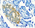
File:Pancreas histology 003.jpg|endocrine (label) | |||
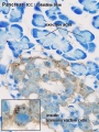
File:Pancreas histology 004.jpg|blood vessels (label) | |||
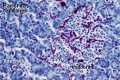
File:Pancreas histology 005.jpg|insulin (label) | |||
File:Pancreas histology 101.jpg|overview | |||
File:Pancreas histology 102.jpg|exocrine | |||
File:Pancreas histology 103.jpg|endocrine | |||
File:Pancreas histology 104.jpg|blood vessels | |||
File:Pancreas histology 105.jpg|insulin | |||
File:Human-_pancreatic_adult_islet.jpg|Islet labeled for insulin and Glucagon | |||
File:Human- pancreatic adult islet-insulin.jpg|Insulin (Fl) | |||
File:Human-_pancreatic_adult_islet-glucagon.jpg|Glucagon (Fl) | |||
</gallery> | |||
{{Pancreas_Histology_Images}} | |||
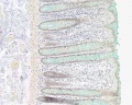
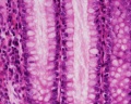
==Intestine== | |||
<gallery> | |||
File:Intestine_villi_crypts_cartoon.jpg|Intestinal villi and crypts cartoon | |||
File:Intestine histology 005.jpg|Duodenum overview | |||
File:Intestine histology 006.jpg|Duodenum villi and crypts | |||
File:Intestine histology 007.jpg|Duodenum | |||
File:Intestine histology 003.jpg|Jejunum overview | |||
File:Intestine histology 004.jpg|Jejunum villus | |||
File:Intestine histology 001.jpg|Jejunum labeled | |||
File:Intestine histology 002.jpg|Jejunum unlabeled | |||
</gallery> | |||
{{Intestine Histology}} | |||
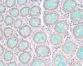
==Colon== | |||
<gallery> | |||
File:Colon histology 006.jpg|Ano-Rectal Junction Overview Labeled | |||
File:Colon histology 001.jpg|Colon Wall Labeled | |||
File:Colon histology 002.jpg|Colon Mucosa Labeled | |||
File:Colon histology 003.jpg|Colon Overview | |||
File:Colon histology 008.jpg|Ano-Rectal Junction Overview | |||
File:Colon histology 007.jpg|Intestinal Gland - longitudinal van Gieson | |||
File:Colon histology 009.jpg|Intestinal Gland - transverse van Gieson | |||
File:Colon histology 004.jpg|Intestinal Gland - longitudinal H&E | |||
File:Colon histology 005.jpg|Intestinal Gland - transverse H&E | |||
</gallery> | |||
{{Colon Histology Links}} | |||
{{Glossary}} | {{Glossary}} | ||
{{Footer}} | {{Footer}} | ||
Latest revision as of 06:41, 21 May 2015
| Embryology - 19 Apr 2024 |
|---|
| Google Translate - select your language from the list shown below (this will open a new external page) |
|
العربية | català | 中文 | 中國傳統的 | français | Deutsche | עִברִית | हिंदी | bahasa Indonesia | italiano | 日本語 | 한국어 | မြန်မာ | Pilipino | Polskie | português | ਪੰਜਾਬੀ ਦੇ | Română | русский | Español | Swahili | Svensk | ไทย | Türkçe | اردو | ייִדיש | Tiếng Việt These external translations are automated and may not be accurate. (More? About Translations) |
Introduction
It would be difficult to show all associated histology on a single page as there are many different components of the gastrointestinal tract and associated organs. Therefore this page should be considered as simply a start page containing image galleries and links to explore more specific topics shown below.
Please note that most of these histology images are adult tissues and may be representative sections from other species.
Opening a specific image from the gallery will provide additional description notes, species, staining and links to associated resources.
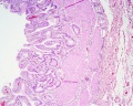
Oesophagus


Stomach
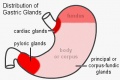
Stomach gastric gland distribution cartoon
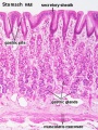
stomach labeled overview
parietal cells - chief cells
mucus neck - parietal cells - chief cells
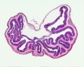
stomach overview
stomach mucosa
mucosa - secretory epithelial sheath - goblet cell
gastric glands - parietal cells - chief cells
stomach overview


- Stomach Histology Links: stomach labeled overview | parietal cells - chief cells | mucus neck - parietal cells - chief cells | stomach overview | stomach mucosa | mucosa - secretory epithelial sheath - goblet cell | gastric glands - parietal cells - chief cells | stomach overview | Stomach Histology | Stomach Development | Gastrointestinal Tract Development
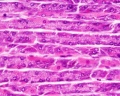
Liver
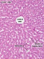
Central vein (label)
Central vein (unlabel)
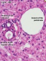
Portal triad 1 (label)
Portal triad 2 (label)

Portal triad (unlabel)
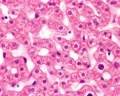
Hepatocytes (unlabel)
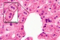
Hepatocytes polyploid (label)


- Liver Histology: Central vein (label) | Central vein (unlabel) | Portal triad 1 (label) | Portal triad 2 (label) | Portal triad (unlabel) | Hepatocytes (unlabel) | Hepatocytes polyploid (label) | Liver - reticular connective tissue (LP) | Liver - reticular connective tissue (HP) | Liver - fetal (HP) | Liver - fetal (HP) | Liver Development | GIT Histology
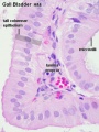
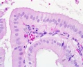
Gall Bladder
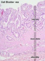
overview (label)
overview (unlabel)
epithelium (label)
epithelium (unlabel)


- Gallbladder Histology: overview (label) | overview (unlabel) | epithelium (label) | epithelium (unlabel) | GIT Histology
Pancreas
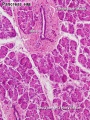
overview (label)
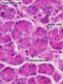
exocrine (label)
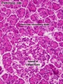
endocrine (label)
blood vessels (label)
insulin (label)
overview
exocrine
endocrine
blood vessels
insulin
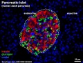
Islet labeled for insulin and Glucagon
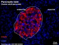
Insulin (Fl)
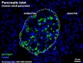
Glucagon (Fl)


- Pancreas Histology Links: overview (label) | exocrine (label) | endocrine (label) | blood vessels (label) | insulin (label) | overview | exocrine | endocrine | blood vessels | insulin | Islet labeled for insulin and Glucagon | Insulin (Fl) | Glucagon (Fl) | GIT Histology
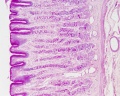
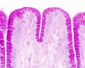
Intestine
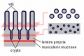
Intestinal villi and crypts cartoon
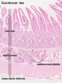
Duodenum overview
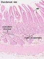
Duodenum villi and crypts
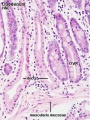
Duodenum
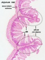
Jejunum overview
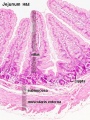
Jejunum villus
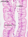
Jejunum labeled
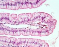
Jejunum unlabeled


- Intestine Histology Links: Duodenum overview | Duodenum villi and crypts | Duodenum | Jejunum overview | Jejunum villus | Jejunum labeled | Jejunum unlabeled | Gastrointestinal Tract Histology | Intestine Development
Colon
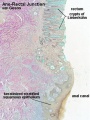
Ano-Rectal Junction Overview Labeled
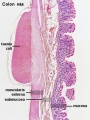
Colon Wall Labeled
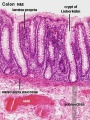
Colon Mucosa Labeled
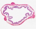
Colon Overview
Ano-Rectal Junction Overview
Intestinal Gland - longitudinal van Gieson
Intestinal Gland - transverse van Gieson
Intestinal Gland - longitudinal H&E
Intestinal Gland - transverse H&E


- Colon Histology Links: Ano-Rectal Junction Overview Labeled | Colon Wall Labeled | Colon Mucosa Labeled | Colon Overview | Ano-Rectal Junction Overview | Intestinal Gland - longitudinal van Gieson | Intestinal Gland - transverse van Gieson | Intestinal Gland - longitudinal H&E | Intestinal Gland - transverse H&E | GIT Histology | Gastrointestinal Tract Development
Glossary Links
- Glossary: A | B | C | D | E | F | G | H | I | J | K | L | M | N | O | P | Q | R | S | T | U | V | W | X | Y | Z | Numbers | Symbols | Term Link
Cite this page: Hill, M.A. (2024, April 19) Embryology Gastrointestinal Tract - Histology. Retrieved from https://embryology.med.unsw.edu.au/embryology/index.php/Gastrointestinal_Tract_-_Histology
- © Dr Mark Hill 2024, UNSW Embryology ISBN: 978 0 7334 2609 4 - UNSW CRICOS Provider Code No. 00098G