Frog Development: Difference between revisions
| Line 33: | Line 33: | ||
| | | | ||
* '''Variation in the schedules of somite and neural development in frogs'''<ref name=PMID23184997><pubmed>23184997</pubmed></ref> "The timing of notochord, somite, and neural development was analyzed in the embryos of six different frog species, which have been divided into two groups, according to their developmental speed. Rapid developing species investigated were Xenopus laevis (Pipidae), Engystomops coloradorum, and Engystomops randi (Leiuperidae). The slow developers were Epipedobates machalilla and Epipedobates tricolor (Dendrobatidae) and Gastrotheca riobambae (Hemiphractidae). ...We propose that these changes are achieved through differential timing of developmental modules that begin with the elongation of the notochord during gastrulation in the rapidly developing species. The differences might be related to the necessity of developing a free-living tadpole quickly in rapid developers." | |||
* '''Unfertilized frog eggs die by apoptosis following meiotic exit'''<ref><pubmed>22195698</pubmed></ref>"Here, we report that the vast majority of naturally laid unfertilized eggs of the African clawed frog Xenopus laevis spontaneously exit metaphase arrest under various environmental conditions and degrade by a well-defined apoptotic process within 48 hours after ovulation. The main features of this process include cytochrome c release, caspase activation, ATP depletion, increase of ADP/ATP ratio, apoptotic nuclear morphology, progressive intracellular acidification, and egg swelling. Meiotic exit seems to be a prerequisite for execution of the apoptotic program." | * '''Unfertilized frog eggs die by apoptosis following meiotic exit'''<ref><pubmed>22195698</pubmed></ref>"Here, we report that the vast majority of naturally laid unfertilized eggs of the African clawed frog Xenopus laevis spontaneously exit metaphase arrest under various environmental conditions and degrade by a well-defined apoptotic process within 48 hours after ovulation. The main features of this process include cytochrome c release, caspase activation, ATP depletion, increase of ADP/ATP ratio, apoptotic nuclear morphology, progressive intracellular acidification, and egg swelling. Meiotic exit seems to be a prerequisite for execution of the apoptotic program." | ||
| Line 41: | Line 43: | ||
* '''Repression of zygotic gene expression in the Xenopus germline.''' <ref><pubmed>20110330</pubmed></ref> "Primordial germ cells (PGCs) in Xenopus are specified through the inheritance of germ plasm. During gastrulation, PGCs remain totipotent while surrounding cells in the vegetal mass become committed to endoderm through the action of the vegetal localized maternal transcription factor VegT. We find that although PGCs contain maternal VegT RNA, they do not express its downstream targets at the mid-blastula transition (MBT)." | * '''Repression of zygotic gene expression in the Xenopus germline.''' <ref><pubmed>20110330</pubmed></ref> "Primordial germ cells (PGCs) in Xenopus are specified through the inheritance of germ plasm. During gastrulation, PGCs remain totipotent while surrounding cells in the vegetal mass become committed to endoderm through the action of the vegetal localized maternal transcription factor VegT. We find that although PGCs contain maternal VegT RNA, they do not express its downstream targets at the mid-blastula transition (MBT)." | ||
|} | |} | ||
Revision as of 21:48, 1 December 2012
Introduction


The frog has been historically been used as an amphibian animal model of development due to the ease of observation from the fertilized egg through to tadpole stage. The later metamorphosis of the tadpole to frog has also been studied for hormonal controls and limb development. There have also been many different species used in these developmental studies.
The frog was historically used by many of the early embryology investigators and currently there are many different molecular mechanisms concerning development of the frog. The 2012 Nobel prize in medicine was recently awarded to John Gurdon for his 1960's experiments involving nuclear transplantation with adult nuclei into frog eggs, these studies were the precursor to current research in stem cells.
The African clawed frog (Xenopus laevis) has been used in many embryological and electrophysiological studies as well as the basis of a historic pregnancy test. The advantages of this frog is the fertility cycle can be easliy controlled and the eggs develop entirely independently and easily visible to the investigator. You can see an overview of the frog life cycle with links to specific stages as well as movies of the early process of gastrulation. This animal model has also shown that localization of maternal messenger RNA (eg vegetal and review) appears to play a key role in the development of early embryological patterns.
The Leopard frog (Rana pipiens) in 1952 became the first successful nuclear transfer experiment. Nuclear transfer is an embryological technique, and involves removal of the nucleus from an egg and replacement with the nucleus of another donor cell. This experiment paved the way for what we know today as the field of cloning.[1]
In Australia, the cane toad (Bufo marinus) species was introduced in 1935 to control cane insect pests. It has now itself become an introduced pest and has also been studied/used more in order to try and biologically control. The area which they occupy has continued to expand. This toad has a poisonous secretion that is extremely toxic and should be handled with care at all times.
| Animal Development: axolotl | bat | cat | chicken | cow | dog | dolphin | echidna | fly | frog | goat | grasshopper | guinea pig | hamster | horse | kangaroo | koala | lizard | medaka | mouse | opossum | pig | platypus | rabbit | rat | salamander | sea squirt | sea urchin | sheep | worm | zebrafish | life cycles | development timetable | development models | K12 |
Some Recent Findings

|
Recent References | References
Taxon
Xenopus Laevis
Eukaryotae; mitochondrial eukaryotes; Metazoa; Chordata;Vertebrata; Amphibia; Batrachia; Anura; Mesobatrachia; Pipoidea;Pipidae; Xenopodinae; Xenopus

Rana pipiens

Taxonomy Id: 8404 Preferred common name: northern leopard frog Rank: species
Genetic code: Translation table 1 (Standard) Mitochondrial genetic code: Translation table 2 Lineage( abbreviated ):
Eukaryota; Metazoa; Chordata; Craniata; Vertebrata; Amphibia; Batrachia; Anura; Neobatrachia; Ranoidea; Ranidae; Raninae; Rana
Frog Life Cycle

Development Timeline
Typical frog development at 18oC from fertilised egg.
- 0 hours - fertilization of the egg
- 1 hours - formation of the gray crescent due to pigment migration
- 3.5 hours - early cleavage
- 4.5 hours - blastula stage(coeloblastula with eccentric blastocoel
- 26 hours - gastrulation
- 26 hours - early crescent shaped dorsal lip
- 34 hours middle semicircular blastoporal lip
- 42 hours late circular blastoporal lip
- 50 hours - neurulation
- 50 hours - early medullary plate
- 62 hours - middle neural folds converging
- 67 hours - late neural tube formed and ciliation of embryo
- 84 hours - tail bud stage(early organogeny)
- 96 hours - muscular response to tactile stimulation
- 118 hours - early heart beat, development of gill buds
- 140 hours - hatching and gill circulation
- 162 hours - mouth opens and cornea becomes transparent
- 192 hours - tail fin circulation established
- 216 hours - degeneration of external gills, formation of operculum, development of embryonic teeth
- 240 hours - opercular fold over brachial chamber except for spiracle and internal gills
- 255 hours - prolonged larval stage with refinement of organs
- 270 hours - development of hindlimbs, internal development of forelimbs in opercular cavity
- 275 hours - projection of forelimbs through operculum, left side first
- 280 hours - absorption of the tail and reduction in size of the gut
- 284 hours - metamorphosis complete, emergence from water as miniature, air breathing frog
Oocyte Balbiani body
- spherical cytoplasmic region that forms within the oocyte in early oogenesis and then fragments and disperses in late oogenesis.
- membrane-less structure consisting of mitochondria, endoplasmic reticulum (ER), membranous vesicles and lipid droplets.
Xenopus stage I oocytes
- Balbiani body is ∼40 μm in diameter
- contains half a million mitochondria, with different morphology and metabolism from other cytoplasmic mitochondria
- rich in membranous vesicles, and ER cysternae.
- vegetal apex (METRO region) contains germinal granules and localized RNAs
- Xlsirts[7]
- family of interspersed repeat RNAs that contain from 3 to 13 repeat units (each 79 to 81 nucleotides long) flanked by unique sequences.
- homologous to the mammalian Xist gene involved in X chromosome inactivation
- stage 2 oocytes - appears first in the mitochondrial cloud (Balbiani body)
- stage 3 oocytes - translocated as island-like structures to the vegetal cortex coincident with the localization of the germ plasm.
Germ Layers
The following paper cartoons[8] show models of signaling mechanisms that occur during early development of the germ cell layers (ectoderm, mesoderm and endoderm).

|

|
References
- ↑ <pubmed>16589125</pubmed>| PMC1063586 | PNAS Classic
- ↑ <pubmed>19549299</pubmed>| PMC2706234 | BMC Dev Biol.
- ↑ <pubmed>23184997</pubmed>
- ↑ <pubmed>22195698</pubmed>
- ↑ <pubmed>21042572</pubmed>
- ↑ <pubmed>20110330</pubmed>
- ↑ <pubmed>7505061</pubmed>
- ↑ <pubmed>17925852</pubmed>| PMC1994590 | PLoS ONE
Search Pubmed: frog development | xenopus development

Additional Images
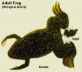
Adult female xenopus laevis with eggs
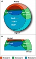
Germ layer signaling
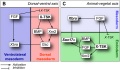
Germ layer signaling

External Links
External Links Notice - The dynamic nature of the internet may mean that some of these listed links may no longer function. If the link no longer works search the web with the link text or name. Links to any external commercial sites are provided for information purposes only and should never be considered an endorsement. UNSW Embryology is provided as an educational resource with no clinical information or commercial affiliation.
- Xenbase A database of information pertaining to the cell and developmental biology of the frog, Xenopus
- Xenopus Laboratory List A database of Labs studying Xenopus
- Xenopus Microarrays
- Xenopus Cell Biology
- The Xenopus Molecular Marker Resource An electronic library of information on embryonic development of the frog, Xenopus laevis | Index page for all Markers | whole mount staining patterns
- Molecular Markers of Development: cement gland XA, XAG, XCG | early mesoderm - BMP2, BMP4, Chordin, goosecoid, Mix,[Marker_pages/organizer/noggin.html noggin], Xbra, Xnr3, Xwnt-8, XVent1 and XVent2 | endothelial - Xl-fli | germ cells - Xpat | heart - cardiac troponin I , XNKX-2.5, XTin1 (XNKX-2.3) | lateral line - tor70, [Marker_pages/CNS/2G9.html 2G9] | muscle - 5A3, 12/101, cardiac actin, XMyf-5, XMyoD | neural crest - Slug, XTwist , xAP2 | notochord - Xnot, tor70 | pronephros - [Marker_pages/pronephros/3G8.html 3G8 ], Wilms' tumor (xWT1), Xlim-1, Xwnt-4 | pronephric duct - 4A6
- Frogs of Greater Brisbane Region (Australia)
- Developmental Biology- Laurie Iten's Serially Sectioned Frog and Chick Embryos
- Developmental Biology- Jeff Hardin's Amphibian Embryology Tutorial
- NIH- Organisms for biomedical research
- Columbia University Kelley Lab - The natural and unnatural histories of xenopus laevis
| Animal Development: axolotl | bat | cat | chicken | cow | dog | dolphin | echidna | fly | frog | goat | grasshopper | guinea pig | hamster | horse | kangaroo | koala | lizard | medaka | mouse | opossum | pig | platypus | rabbit | rat | salamander | sea squirt | sea urchin | sheep | worm | zebrafish | life cycles | development timetable | development models | K12 |
Glossary Links
- Glossary: A | B | C | D | E | F | G | H | I | J | K | L | M | N | O | P | Q | R | S | T | U | V | W | X | Y | Z | Numbers | Symbols | Term Link
Cite this page: Hill, M.A. (2024, April 25) Embryology Frog Development. Retrieved from https://embryology.med.unsw.edu.au/embryology/index.php/Frog_Development
- © Dr Mark Hill 2024, UNSW Embryology ISBN: 978 0 7334 2609 4 - UNSW CRICOS Provider Code No. 00098G