File:Zygote - this image.png
Zygote_-_this_image.png (372 × 166 pixels, file size: 83 KB, MIME type: image/png)
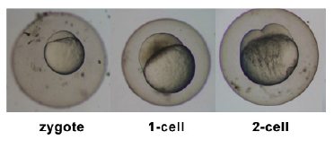
Zebrafish Zygote
- Links: zebrafish
Image Source: Image kindly contributed by Dr Judith Cebra-Thomas Department of Biology, Millersville University. From her Lab Protocols showing Zebrafish Stages sequence based on previously published sources including; Kimmel CB, Ballard WW, Kimmel SR, Ullmann B & Schilling TF. (1995). Stages of embryonic development of the zebrafish. Dev. Dyn. , 203, 253-310. PMID: 8589427 DOI.
- Note - This image was originally uploaded as part of an undergraduate science student project and may contain inaccuracies in either description or acknowledgements. Students have been advised in writing concerning the reuse of content and may accidentally have misunderstood the original terms of use. If image reuse on this non-commercial educational site infringes your existing copyright, please contact the site editor for immediate removal.
Cite this page: Hill, M.A. (2024, April 18) Embryology Zygote - this image.png. Retrieved from https://embryology.med.unsw.edu.au/embryology/index.php/File:Zygote_-_this_image.png
- © Dr Mark Hill 2024, UNSW Embryology ISBN: 978 0 7334 2609 4 - UNSW CRICOS Provider Code No. 00098G
File history
Click on a date/time to view the file as it appeared at that time.
| Date/Time | Thumbnail | Dimensions | User | Comment | |
|---|---|---|---|---|---|
| current | 17:08, 20 September 2009 |  | 372 × 166 (83 KB) | Z3218657 (talk | contribs) | This image has been provide by by Judy Cebra-Thomas [http://www.swarthmore.edu/NatSci/sgilber1/DB_lab/Fish/fish_stage.html] |
| 16:42, 20 September 2009 |  | 372 × 166 (83 KB) | Z3218657 (talk | contribs) |
You cannot overwrite this file.
File usage
The following 5 pages use this file:
