File:ZPulmonary Atresia.jpg
Original file (653 × 618 pixels, file size: 85 KB, MIME type: image/jpeg)
What am I looking at?
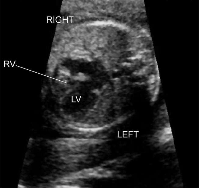
This is a 2D ultrasound of a fetal heart, showing pulmonary atresia with in intact ventricular septum. This is a four-chamber view of the heart showing that the left ventricle is greater in size than the right ventricle. The right ventricle is hypoplastic, and the right ventricular cavity is small.
Image Copyright Information
Image sourced from:[1]
Article: <pubmed>PMC2840777</pubmed>
Image copyright statement:
This is an open-access article distributed under the terms of the Creative Commons Attribution License, which permits unrestricted use, distribution, and reproduction in any medium, provided the original work is properly cited.
File history
Click on a date/time to view the file as it appeared at that time.
| Date/Time | Thumbnail | Dimensions | User | Comment | |
|---|---|---|---|---|---|
| current | 17:07, 26 September 2010 | | 653 × 618 (85 KB) | Z3252833 (talk | contribs) | ==What am I looking at?== This is a 2D ultrasound of a fetal heart, showing pulmonary atresia with in intact ventricular septum. This is a four-chamber view of the heart showing that the left ventricle is greater in size than the right ventricle. The rig |
You cannot overwrite this file.
File usage
The following page uses this file: