File:West11.jpg

Original file (367 × 963 pixels, file size: 27 KB, MIME type: image/jpeg)
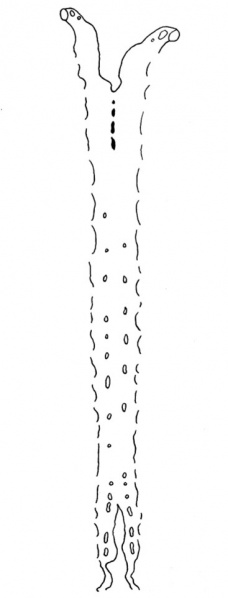
Fig. 11. General plan of the Aorta
The general plan of the aorta can be seen in the figure (11). It passes cranial wards from the bulbus and then bifurcates, and the two divisions turn round in a caudal direction to form the first pair of aortic arches, and from the summit of each there arises the internal carotid artery. There is a second pair of aortic arches caudal to the first,and a third pair is almost completed by a considerable outgrowth from the ventral aorta and a much smaller ventral sprout from the dorsal aorta. The dorsal aortae then pass on along each side of the middle line as far as the caudal border of the 5th somite, or the junction of brain and spinal cord, where they join here and there; but true union does not occur til the level of the middle of the 7th somite,or at the level of the cranial end of the liver;from this point a single dorsal aorta passes towards the tail as far as the origin of the allantois, at the level of the 20th somite, where a division into two caudal aortae takes place, and here too the large umbilical plexus of vessels arises. The condition is thus rather in advance of that found in Girgis' (1926) embryo of 22 somites,and is very like that in Johnson's (1917) specimen of 24 somites.
Ventral branches, not strictly segmentally arranged, arise from the ventral surface of the aorta; there are 12 on the left side and 14 on the right. Dorsal and lateral branches are present also, but are not so definite as the ventral series. The picture formed by these various branches of the aorta and by the post-cardinal and sub-cardinal veins is almost identical with H. M. Evans' (1912) Fig. 436, p. 632, of the vessels in an embryo of 23 somites.
The more caudal of the ventral branches of the aorta, which rise in between the 20th and 23rd somites, i.e. between the 9th and 12th thoracic segments, are those which give origin to the umbilical arteries; these two vessels run, one on each side of the allantois as it leaves the gut, but ventral and dorsal, the left being ventral, to the allantois as itliesinthebodystalk; a branch connects the two arteries round the left side of the allantois just before its expanded end, and that is the only communication between the two vessels; the umbilical arteries then break up into branches in the chorion.
| Historic Disclaimer - information about historic embryology pages |
|---|
| Pages where the terms "Historic" (textbooks, papers, people, recommendations) appear on this site, and sections within pages where this disclaimer appears, indicate that the content and scientific understanding are specific to the time of publication. This means that while some scientific descriptions are still accurate, the terminology and interpretation of the developmental mechanisms reflect the understanding at the time of original publication and those of the preceding periods, these terms, interpretations and recommendations may not reflect our current scientific understanding. (More? Embryology History | Historic Embryology Papers) |
- Links: Fig 1 | Fig 2 | Fig 3 | Fig 4 | Fig 5 | Fig 6 | Fig 7 | Fig 8 | Fig 9 | Fig 10 | Fig 11 | Plate 1 | Plate 1 Fig 1 | Plate 1 Fig 2 | Plate 1 Fig 3 | Plate 1 Fig 4 | Plate 1 Fig 5 | Plate 1 Fig 6
Reference
West CM. A human embryo of twenty-five somites. (1937) J. Anat., 71(2): 169-200.1. PMID 17104635
Cite this page: Hill, M.A. (2024, April 19) Embryology West11.jpg. Retrieved from https://embryology.med.unsw.edu.au/embryology/index.php/File:West11.jpg
- © Dr Mark Hill 2024, UNSW Embryology ISBN: 978 0 7334 2609 4 - UNSW CRICOS Provider Code No. 00098G
File history
Click on a date/time to view the file as it appeared at that time.
| Date/Time | Thumbnail | Dimensions | User | Comment | |
|---|---|---|---|---|---|
| current | 15:23, 28 January 2012 | 367 × 963 (27 KB) | S8600021 (talk | contribs) | ==Fig. 11 == {{Template:West1937}} {{Historic Disclaimer}} {{Historic Papers}} ===Reference=== <pubmed>17104635</pubmed>| [http://www.ncbi.nlm.nih.gov/pmc/articles/PMC1252340 PMC1252340] Category:Carnegie Stage 12 |
You cannot overwrite this file.
File usage
The following page uses this file:
