File:West10.jpg: Difference between revisions
No edit summary |
mNo edit summary |
||
| Line 1: | Line 1: | ||
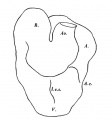
==Fig. 10 == | ==Fig. 10. Drawing of a Heart Model== | ||
As can be seen from the drawing of a model (Text-fig. 10), the heart consists of (1) a sinus venosus, in the upper left part of which is the opening into (2) the atrium which is, for the most part, a large single chamber, but which in its caudal part shows by an external groove and an internal ridge a commencing division into right and left chambers. The atrium leads by a constricted canal into (3) the ventricle, which forms the most caudal part of the heart and also shows the beginning of an interventricular septum and which then turns obliquely cranialwards and to the right; at the summit of its arch the ventricle joins (4) the bulbus, which passes caudalwards and to the left, extending from the most cranial limit of the heart to about half-way down it, where it turns abruptly cranialwards into (5) the aorta. The sharpness of the various curvatures is emphasized more in the endothelial heart tube than in the myocardial. The caudal part of the sinus venosus lies in the septum transversum and is slung for some distance to the cranial side of its union with the atrium to the middorsal line by a thick mesentery which is common to the heart and the gut, the heart lying ventral to the lung bud region, and the two organs being separated from each other only by the thickness of the irrespective walls. A mesentery slings the arterial end of the heart to the ventral body wall for a short distance until the aorta becomes embedded in the body wall. Elsewhere the heart tube is free. | |||
With regard to structure; the sinus venosus is different from the rest of the heart and has not the same thick myocardial wall, but is more of the nature of a largely dilated vessel, it is indeed a true venous sinus. | |||
In those parts of the heart where the walls are thin there is a considerable interval between the endocardial and myocardial heart tubes, which is filed with the loose trabecular tissue which has been so often described. Inthe thickest part of the heart, e.g. the ventricle, such a submyocardial space scarcely exists owing to the thickness of the muscular wall of the heart, and here the endocardial tube is formed of extremely thin, elongated cells placed endtoend;where there is a large sub myocardial space it can be seen that the trabecular tissue forms, on the deep surface of the space, a definite limiting membrane which abuts against the endocardial tube. | |||
There is a difference in the appearance of the myocardium in the various parts of the heart; the sinus is by far the thinnest, and really has no myocardial coattail; a myocardial coat can be recognised in the atrium, where it forms a fairly thick and compact layer of cels,quite different from what is found in the wall of the ventricle; in the ventricle the myocardium is thicker but the cells are more loosely arranged, they are large and form columns or chains enclosing clear spaces between them; the myocardium of the bulbus forms a layer intermediate in thickness between that of the sinus and the atrium. | |||
{{Historic | {{Historic Disclaimer}} | ||
{{West1937}} | |||
===Reference=== | ===Reference=== | ||
West, CM. [[Paper - A Human Embryo of Twenty-five Somites|'''A Human Embryo of Twenty-five Somites''']]. J. Anat.: 1937, 71(Pt 2);169-200.1 PMID 17104635 | |||
{{Footer}} | |||
[[Category: | [[Category:Carnegie Stage 12]] | ||
[[Category:Heart]] | |||
Revision as of 12:21, 17 February 2016
Fig. 10. Drawing of a Heart Model
As can be seen from the drawing of a model (Text-fig. 10), the heart consists of (1) a sinus venosus, in the upper left part of which is the opening into (2) the atrium which is, for the most part, a large single chamber, but which in its caudal part shows by an external groove and an internal ridge a commencing division into right and left chambers. The atrium leads by a constricted canal into (3) the ventricle, which forms the most caudal part of the heart and also shows the beginning of an interventricular septum and which then turns obliquely cranialwards and to the right; at the summit of its arch the ventricle joins (4) the bulbus, which passes caudalwards and to the left, extending from the most cranial limit of the heart to about half-way down it, where it turns abruptly cranialwards into (5) the aorta. The sharpness of the various curvatures is emphasized more in the endothelial heart tube than in the myocardial. The caudal part of the sinus venosus lies in the septum transversum and is slung for some distance to the cranial side of its union with the atrium to the middorsal line by a thick mesentery which is common to the heart and the gut, the heart lying ventral to the lung bud region, and the two organs being separated from each other only by the thickness of the irrespective walls. A mesentery slings the arterial end of the heart to the ventral body wall for a short distance until the aorta becomes embedded in the body wall. Elsewhere the heart tube is free.
With regard to structure; the sinus venosus is different from the rest of the heart and has not the same thick myocardial wall, but is more of the nature of a largely dilated vessel, it is indeed a true venous sinus.
In those parts of the heart where the walls are thin there is a considerable interval between the endocardial and myocardial heart tubes, which is filed with the loose trabecular tissue which has been so often described. Inthe thickest part of the heart, e.g. the ventricle, such a submyocardial space scarcely exists owing to the thickness of the muscular wall of the heart, and here the endocardial tube is formed of extremely thin, elongated cells placed endtoend;where there is a large sub myocardial space it can be seen that the trabecular tissue forms, on the deep surface of the space, a definite limiting membrane which abuts against the endocardial tube.
There is a difference in the appearance of the myocardium in the various parts of the heart; the sinus is by far the thinnest, and really has no myocardial coattail; a myocardial coat can be recognised in the atrium, where it forms a fairly thick and compact layer of cels,quite different from what is found in the wall of the ventricle; in the ventricle the myocardium is thicker but the cells are more loosely arranged, they are large and form columns or chains enclosing clear spaces between them; the myocardium of the bulbus forms a layer intermediate in thickness between that of the sinus and the atrium.
| Historic Disclaimer - information about historic embryology pages |
|---|
| Pages where the terms "Historic" (textbooks, papers, people, recommendations) appear on this site, and sections within pages where this disclaimer appears, indicate that the content and scientific understanding are specific to the time of publication. This means that while some scientific descriptions are still accurate, the terminology and interpretation of the developmental mechanisms reflect the understanding at the time of original publication and those of the preceding periods, these terms, interpretations and recommendations may not reflect our current scientific understanding. (More? Embryology History | Historic Embryology Papers) |
- Links: Fig 1 | Fig 2 | Fig 3 | Fig 4 | Fig 5 | Fig 6 | Fig 7 | Fig 8 | Fig 9 | Fig 10 | Fig 11 | Plate 1 | Plate 1 Fig 1 | Plate 1 Fig 2 | Plate 1 Fig 3 | Plate 1 Fig 4 | Plate 1 Fig 5 | Plate 1 Fig 6
Reference
West CM. A human embryo of twenty-five somites. (1937) J. Anat., 71(2): 169-200.1. PMID 17104635
Cite this page: Hill, M.A. (2024, April 19) Embryology West10.jpg. Retrieved from https://embryology.med.unsw.edu.au/embryology/index.php/File:West10.jpg
- © Dr Mark Hill 2024, UNSW Embryology ISBN: 978 0 7334 2609 4 - UNSW CRICOS Provider Code No. 00098G
Reference
West, CM. A Human Embryo of Twenty-five Somites. J. Anat.: 1937, 71(Pt 2);169-200.1 PMID 17104635
Cite this page: Hill, M.A. (2024, April 19) Embryology West10.jpg. Retrieved from https://embryology.med.unsw.edu.au/embryology/index.php/File:West10.jpg
- © Dr Mark Hill 2024, UNSW Embryology ISBN: 978 0 7334 2609 4 - UNSW CRICOS Provider Code No. 00098G
File history
Click on a date/time to view the file as it appeared at that time.
| Date/Time | Thumbnail | Dimensions | User | Comment | |
|---|---|---|---|---|---|
| current | 15:23, 28 January 2012 |  | 501 × 539 (25 KB) | S8600021 (talk | contribs) | ==Fig. 10 == {{Template:West1937}} {{Historic Disclaimer}} {{Historic Papers}} ===Reference=== <pubmed>17104635</pubmed>| [http://www.ncbi.nlm.nih.gov/pmc/articles/PMC1252340 PMC1252340] Category:Carnegie Stage 12 |
You cannot overwrite this file.
File usage
The following page uses this file:
