File:Waterston20.jpg
From Embryology

Size of this preview: 416 × 599 pixels. Other resolution: 500 × 720 pixels.
Original file (500 × 720 pixels, file size: 75 KB, MIME type: image/jpeg)
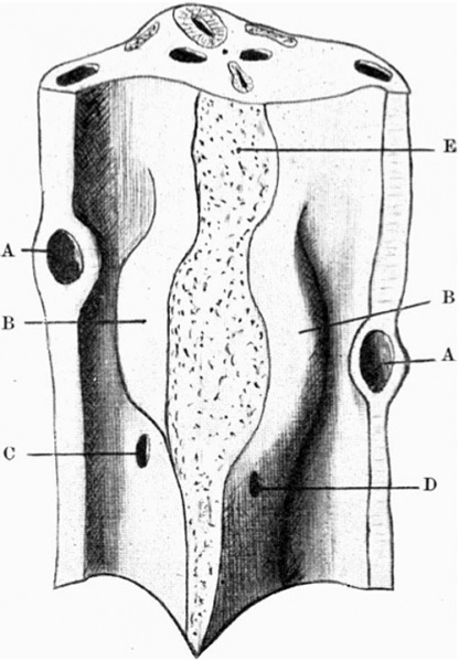
Fig. 20. Model after removal of the septum transversum.
Wax-plate reconstruction model in fig.19, after removal of the septum transversum, showing the dorsal wall ofpericardial and peritoneal ccelom with orifices ofright and left pneumato-enteric recesses.
The dotted area indicates, above, the attachment of the dorsal mesocardium, and, below, the ventral mesentery.
Legend
- A, ducts of Cuvier
- B, mesodermal lung thickenings; orifices of pneumato-enteric recesses
Carnegie Staging Comparison: A 27 somite stage embryo would be similar to a Carnegie stage 12 (26 - 30 days), caudal neuropore closes, Somite Number 21-29.
| Historic Disclaimer - information about historic embryology pages |
|---|
 |
- Historic Paper Links: 13-14 Somites | 22 Somites | 23 Somites | 25 Somites | 27 Somites | Mall Human Embryo Collection | Embryology History | Carnegie stage 11 | Carnegie stage 12 | Journal of Anatomy | Embryonic Development | Category:Historic Embryology
Reference
<pubmed>17233016</pubmed>| PMC1288995
File history
Click on a date/time to view the file as it appeared at that time.
| Date/Time | Thumbnail | Dimensions | User | Comment | |
|---|---|---|---|---|---|
| current | 13:56, 27 January 2012 | | 500 × 720 (75 KB) | S8600021 (talk | contribs) | ==Fig. 20. Model after removal of the septum transversum.== Wax-plate reconstruction model in fig.19, after removal of the septum transversum, showing the dorsal wall ofpericardial and peritoneal ccelom with orifices ofright and left pneumato-enteric rec |
You cannot overwrite this file.