File:Waterston13.jpg
From Embryology
Size of this preview: 377 × 598 pixels. Other resolution: 429 × 681 pixels.
Original file (429 × 681 pixels, file size: 63 KB, MIME type: image/jpeg)
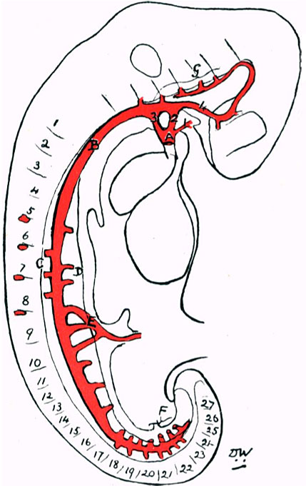
Fig. 13. Linear profile reconstruction of arteries
Legend
- A, conus arteriosus
- B, dorsal aorta
- C, dorsal segmental vessels
- D, ventral segmental vessels
- E, vitelline rootlets
- F, umbilical rootlets
- G, segmental vessels to hindbrain.
Carnegie Staging Comparison: A 27 somite stage embryo would be similar to a Carnegie stage 12 (26 - 30 days), caudal neuropore closes, Somite Number 21-29.
| Historic Disclaimer - information about historic embryology pages |
|---|
 |
- Historic Paper Links: 13-14 Somites | 22 Somites | 23 Somites | 25 Somites | 27 Somites | Mall Human Embryo Collection | Embryology History | Carnegie stage 11 | Carnegie stage 12 | Journal of Anatomy | Embryonic Development | Category:Historic Embryology
Reference
<pubmed>17233016</pubmed>| PMC1288995
File history
Click on a date/time to view the file as it appeared at that time.
| Date/Time | Thumbnail | Dimensions | User | Comment | |
|---|---|---|---|---|---|
| current | 12:52, 27 January 2012 | | 429 × 681 (63 KB) | S8600021 (talk | contribs) | <pubmed>17233016</pubmed>| [http://www.ncbi.nlm.nih.gov/pmc/articles/PMC1288995 PMC1288995] ===Historic Embryology=== This is a slightly edited version of the original 1914 paper published in Journal of Anatomy and Physiology. The full paper is still av |
You cannot overwrite this file.