File:Ventricular Septal Defect (VSD).jpeg
.jpeg)
.jpeg&diff=cur&oldid=312962)
.jpeg&direction=next&oldid=312962)
.jpeg&diff=next&oldid=312962)
Ventricular_Septal_Defect_(VSD).jpeg (358 × 402 pixels, file size: 46 KB, MIME type: image/jpeg)
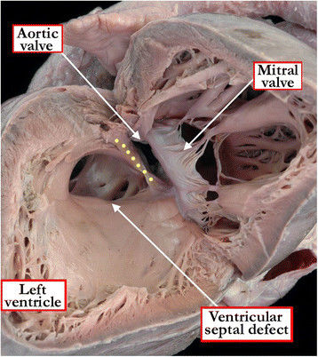
The view from the left ventricle in this specimen shows a ventricular septal defect with exclusively muscular borders opening towards the outlet of the right ventricle, but with postero-caudal deviation of the muscular outlet septum (yellow dots), causing subaortic stenosis.
Reference Diane E Spicer, Hao H Hsu, Jennifer Co-Vu, Robert H Anderson, F Jay Fricker Ventricular septal defect. Orphanet J Rare Dis: 2014, 9;144
The open access articles published in BioMed Central's journals are made available under the Creative Commons Attribution (CC-BY) license, which means they are accessible online without any restrictions and can be re-used in any way, subject only to proper attribution (which, in an academic context, usually means citation).
File history
Click on a date/time to view the file as it appeared at that time.
| Date/Time | Thumbnail | Dimensions | User | Comment | |
|---|---|---|---|---|---|
| current | 13:43, 21 October 2017 | | 358 × 402 (46 KB) | Z5059996 (talk | contribs) | The view from the left ventricle in this specimen shows a ventricular septal defect with exclusively muscular borders opening towards the outlet of the right ventricle, but with postero-caudal deviation of the muscular outlet septum (yellow dots), caus... |
You cannot overwrite this file.
File usage
The following 2 pages use this file:
.jpeg&oldid=312962)