File:Vegf-protects-vessels.JPG: Difference between revisions
(Astrocyte-derived VEGF protects vessels from hyperoxia. After hyperoxia exposure from P7–12 immunohistochemistry was used to visualize vessels with isolectin B4 (green, A, B), collagen IV (green, D, E) and retinal astrocytes (GFAP, red, D, E). (A–C) ) |
No edit summary |
||
| Line 1: | Line 1: | ||
Astrocyte-derived VEGF protects vessels from hyperoxia. | '''Astrocyte-derived VEGF protects vessels from hyperoxia.''' | ||
After hyperoxia exposure from P7–12 immunohistochemistry was used to visualize vessels with isolectin B4 (green, A, B), collagen IV (green, D, E) and retinal astrocytes (GFAP, red, D, E). (A–C) Deletion of astrocyte specific VEGF increased the vaso-obliterated area and led to decreased survival of radial arteries and veins. In some instances this lead to non-perfused, hyperproliferating capillary beds in the periphery (arrowhead in B). (D, E) Retinal astrocyte survival was not affected at this age but in areas of capillary loss astrocytes re-aligned with nerve bundles (arrowheads D, E). Scale bars are 100µm in A and 100µm in D; ** is p<0.01. | After hyperoxia exposure from P7–12 immunohistochemistry was used to visualize vessels with isolectin B4 (green, A, B), collagen IV (green, D, E) and retinal astrocytes (GFAP, red, D, E). (A–C) Deletion of astrocyte specific VEGF increased the vaso-obliterated area and led to decreased survival of radial arteries and veins. In some instances this lead to non-perfused, hyperproliferating capillary beds in the periphery (arrowhead in B). (D, E) Retinal astrocyte survival was not affected at this age but in areas of capillary loss astrocytes re-aligned with nerve bundles (arrowheads D, E). Scale bars are 100µm in A and 100µm in D; ** is p<0.01. | ||
Revision as of 06:04, 3 October 2012
Astrocyte-derived VEGF protects vessels from hyperoxia.
After hyperoxia exposure from P7–12 immunohistochemistry was used to visualize vessels with isolectin B4 (green, A, B), collagen IV (green, D, E) and retinal astrocytes (GFAP, red, D, E). (A–C) Deletion of astrocyte specific VEGF increased the vaso-obliterated area and led to decreased survival of radial arteries and veins. In some instances this lead to non-perfused, hyperproliferating capillary beds in the periphery (arrowhead in B). (D, E) Retinal astrocyte survival was not affected at this age but in areas of capillary loss astrocytes re-aligned with nerve bundles (arrowheads D, E). Scale bars are 100µm in A and 100µm in D; ** is p<0.01.
doi:10.1371/journal.pone.0011863.g003
Source: <pubmed>20686684</pubmed>
Copyright: © 2010 Scott et al. This is an open-access article distributed under the terms of the Creative Commons Attribution License, which permits unrestricted use, distribution, and reproduction in any medium, provided the original author and source are credited.
- Note - This image was originally uploaded as part of an undergraduate science student project and may contain inaccuracies in either description or acknowledgements. Students have been advised in writing concerning the reuse of content and may accidentally have misunderstood the original terms of use. If image reuse on this non-commercial educational site infringes your existing copyright, please contact the site editor for immediate removal.
File history
Click on a date/time to view the file as it appeared at that time.
| Date/Time | Thumbnail | Dimensions | User | Comment | |
|---|---|---|---|---|---|
| current | 06:03, 3 October 2012 | 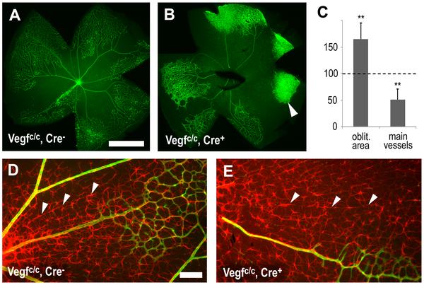 | 600 × 403 (50 KB) | Z3370664 (talk | contribs) | Astrocyte-derived VEGF protects vessels from hyperoxia. After hyperoxia exposure from P7–12 immunohistochemistry was used to visualize vessels with isolectin B4 (green, A, B), collagen IV (green, D, E) and retinal astrocytes (GFAP, red, D, E). (A–C) |
You cannot overwrite this file.
File usage
The following page uses this file: