File:Uveal Melanoma.gif: Difference between revisions
From Embryology
No edit summary |
No edit summary |
||
| Line 1: | Line 1: | ||
[[File:Uveal Melanoma.gif|400px]] | |||
Figure 5. | |||
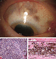
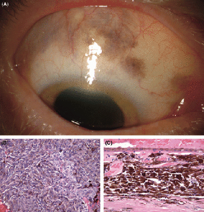
Ciliochoroidal melanoma in an eye with melanocytosis. (A) Diffuse scleral melanocytosis. (B) Photomicrograph demonstrating uveal melanoma, epithelioid‐rich mixed cell type. (Haematoxylin and eosin [H&E]; original magnification [OM] × 100.) (C) Photomicrograph demonstrating adjacent area of choroidal melanocytosis. (H&E; OM × 100.) | |||
===Reference=== | |||
{{#pmid:18547285}} | |||
===Copyright=== | |||
Journal compilation © 2009 Acta Ophthalmol | |||
{{Template:2018 Student Image}} | |||
Latest revision as of 17:12, 12 October 2018
Figure 5.
Ciliochoroidal melanoma in an eye with melanocytosis. (A) Diffuse scleral melanocytosis. (B) Photomicrograph demonstrating uveal melanoma, epithelioid‐rich mixed cell type. (Haematoxylin and eosin [H&E]; original magnification [OM] × 100.) (C) Photomicrograph demonstrating adjacent area of choroidal melanocytosis. (H&E; OM × 100.)
Reference
Horgan N, Shields CL, Swanson L, Teixeira LF, Eagle RC, Ganguly A & Shields JA. (2009). Altered chromosome expression of uveal melanoma in the setting of melanocytosis. Acta Ophthalmol , 87, 578-80. PMID: 18547285 DOI.
Copyright
Journal compilation © 2009 Acta Ophthalmol
- Note - This image was originally uploaded as part of an undergraduate science student 2018 project and may contain inaccuracies in either description or acknowledgements. Students have been advised in writing concerning the reuse of content and may accidentally have misunderstood the original terms of use. If image reuse on this non-commercial educational site infringes your existing copyright, please contact the site editor for immediate removal.
File history
Click on a date/time to view the file as it appeared at that time.
| Date/Time | Thumbnail | Dimensions | User | Comment | |
|---|---|---|---|---|---|
| current | 17:08, 12 October 2018 |  | 480 × 499 (169 KB) | Z5165679 (talk | contribs) |
You cannot overwrite this file.
File usage
The following 3 pages use this file:
