File:UBE3A colocalizes with ASPM at the centrosome throughout mitosis.jpg

Original file (430 × 658 pixels, file size: 130 KB, MIME type: image/jpeg)
UBE3A colocalizes with ASPM at the centrosome throughout mitosis
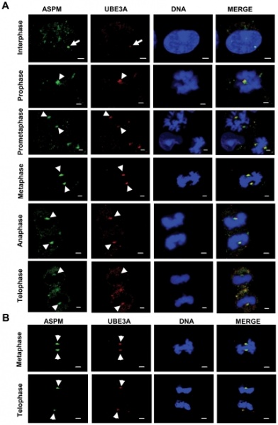
UBE3A colocalizes with ASPM at the centrosome. (A) Indirect immunofluorescence of HEK293 cells at interphase and different phases of mitosis stained with antibodies against ASPM and UBE3A (anti-UBE3A-sc-8926). Note colocalization of UBE3A with ASPM at the centrosome throughout mitosis (arrowheads). Note weak centrosomal staining of UBE3A in an interphase cell (arrow). (B) Indirect immunofluorescence of A549 cells stained with antibodies against UBE3A (anti-UBE3A-sc-8926) and ASPM at metaphase and telophase. Note colocalization of UBE3A with ASPM at the centrosome (arrowheads). Scale bar=2 µm.
Original file name: Figure 3 pone.0020397.g003.jpg
References
<pubmed>21633703</pubmed>| PMC:3102111 | PLoS One
Copyright Singhmar, Kumar. This is an open-access article distributed under the terms of the Creative Commons Attribution License, which permits unrestricted use, distribution, and reproduction in any medium, provided the original author and source are credited.
File history
Click on a date/time to view the file as it appeared at that time.
| Date/Time | Thumbnail | Dimensions | User | Comment | |
|---|---|---|---|---|---|
| current | 11:13, 15 August 2011 |  | 430 × 658 (130 KB) | Z3291643 (talk | contribs) | Pone_0020397_g003.jpg UBE3A colocalizes with ASPM at the centrosome. (A) Indirect immunofluorescence of HEK293 cells at interphase and different phases of mitosis stained with antibodies against ASPM and UBE3A (anti-UBE3A-sc-8926). Note colocalization of |
You cannot overwrite this file.
File usage
The following page uses this file: