File:Trigeminal artery 02.jpg: Difference between revisions
No edit summary |
mNo edit summary |
||
| Line 1: | Line 1: | ||
==Trigeminal artery== | |||
The most common persistent embryonic carotid-vertebrobasilar anastomosis is the trigeminal artery. A persistent trigeminal artery consists of a connection between the intracranial internal carotid artery and the basilar artery, usually in the basilar midsegment or distally near the origin of the superior cerebellar arteries. | |||
:'''Links:''' [[:File:Cerebral brain artery development 01.jpg|Overview cartoon]] | [[:File:Cerebral brain artery development 01.jpg|Early vascular changes]] | [[Cardiovascular System Development]] | [[Neural - Cerebrum Development]] | |||
===Reference=== | |||
<pubmed>26060802</pubmed> | |||
====Copyright==== | |||
© 2015 Korean Stroke Society | |||
(open-access, http://creativecommons.org/licenses/by-nc/3.0/): | |||
This is an Open Access article distributed under the terms of the Creative Commons Attribution Non-Commercial License (http://creativecommons.org/licenses/by-nc/3.0/) which permits unrestricted non-commercial use, distribution, and reproduction in any medium, provided the original work is properly cited. | |||
Jos-17-144-g005.jpg Panel A cropped, resized and labelled from fig. 5. | |||
{{Footer}} | |||
Revision as of 09:39, 5 November 2015
Trigeminal artery
The most common persistent embryonic carotid-vertebrobasilar anastomosis is the trigeminal artery. A persistent trigeminal artery consists of a connection between the intracranial internal carotid artery and the basilar artery, usually in the basilar midsegment or distally near the origin of the superior cerebellar arteries.
- Links: Overview cartoon | Early vascular changes | Cardiovascular System Development | Neural - Cerebrum Development
Reference
<pubmed>26060802</pubmed>
Copyright
© 2015 Korean Stroke Society (open-access, http://creativecommons.org/licenses/by-nc/3.0/): This is an Open Access article distributed under the terms of the Creative Commons Attribution Non-Commercial License (http://creativecommons.org/licenses/by-nc/3.0/) which permits unrestricted non-commercial use, distribution, and reproduction in any medium, provided the original work is properly cited.
Jos-17-144-g005.jpg Panel A cropped, resized and labelled from fig. 5.
Cite this page: Hill, M.A. (2024, April 24) Embryology Trigeminal artery 02.jpg. Retrieved from https://embryology.med.unsw.edu.au/embryology/index.php/File:Trigeminal_artery_02.jpg
- © Dr Mark Hill 2024, UNSW Embryology ISBN: 978 0 7334 2609 4 - UNSW CRICOS Provider Code No. 00098G
File history
Click on a date/time to view the file as it appeared at that time.
| Date/Time | Thumbnail | Dimensions | User | Comment | |
|---|---|---|---|---|---|
| current | 09:38, 5 November 2015 | 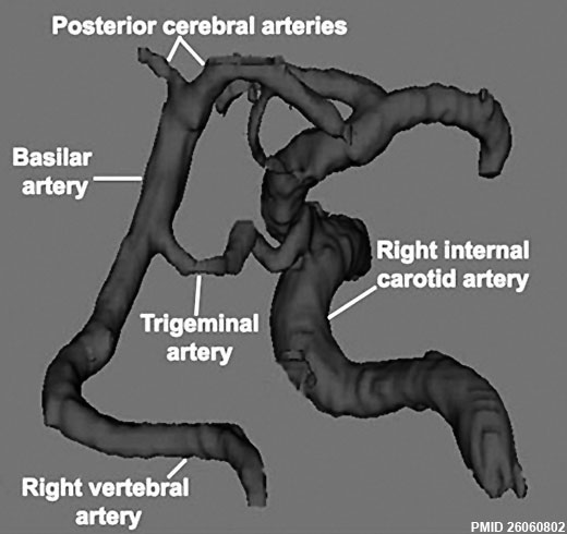 | 520 × 490 (35 KB) | Z8600021 (talk | contribs) |
You cannot overwrite this file.
File usage
The following page uses this file: