File:Thyng1914 plate3b.jpg
From Embryology
Size of this preview: 764 × 600 pixels. Other resolution: 1,911 × 1,500 pixels.
Original file (1,911 × 1,500 pixels, file size: 489 KB, MIME type: image/jpeg)
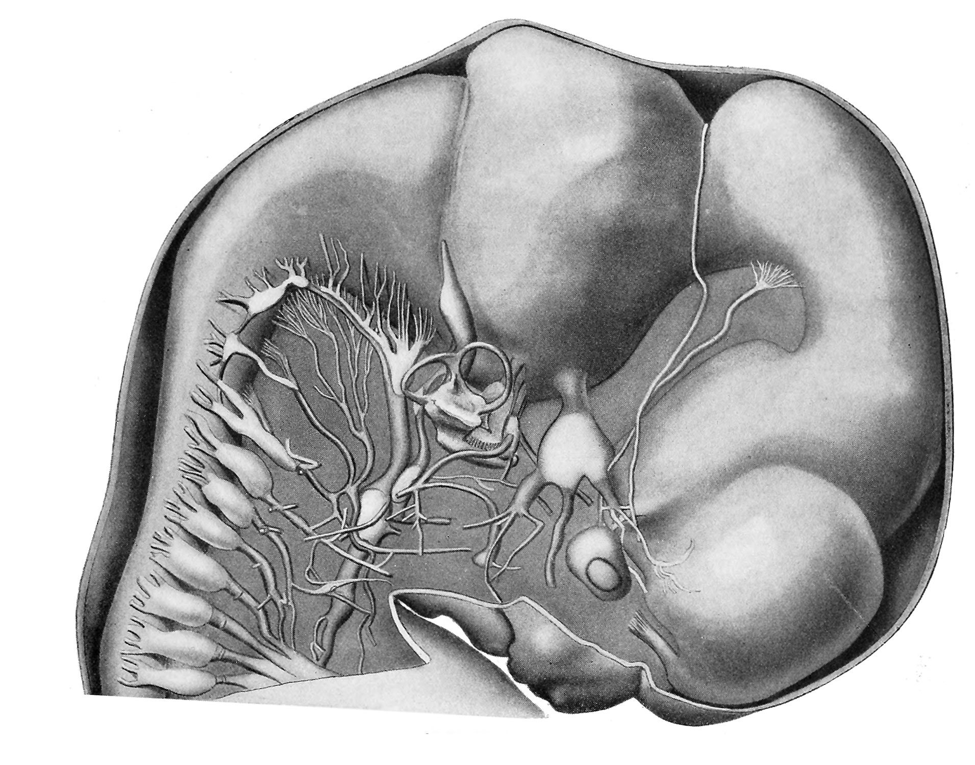
Plate 3. Brain and Cervical Spinal Cord
Reconstruction showing the right side of the brain and the cervical cord ; the right cranial and cervical nerves; the internal ear, and the optic vesicle of a 17.8 mm. human embryo (H. E. C. 839). X 11.9 diams.
- Embryo 17.8 mm Links: Fig 1 | Fig 2 | Plate 1a | Plate 1b | Plate 2a | Plate 2b | Plate 3a | Plate 3b | Plate 4a | Plate 4b | Plate 5a | Plate 5b | Plate 6 | Harvard Collection | Carnegie stage 19
Reference
Thyng FW. The anatomy of a 17.8 mm human embryo. (1914) Amer. J Anat. 17: 31-112.
Cite this page: Hill, M.A. (2024, April 18) Embryology Thyng1914 plate3b.jpg. Retrieved from https://embryology.med.unsw.edu.au/embryology/index.php/File:Thyng1914_plate3b.jpg
- © Dr Mark Hill 2024, UNSW Embryology ISBN: 978 0 7334 2609 4 - UNSW CRICOS Provider Code No. 00098G
File history
Click on a date/time to view the file as it appeared at that time.
| Date/Time | Thumbnail | Dimensions | User | Comment | |
|---|---|---|---|---|---|
| current | 06:41, 10 April 2014 | | 1,911 × 1,500 (489 KB) | Z8600021 (talk | contribs) | |
| 06:40, 10 April 2014 | 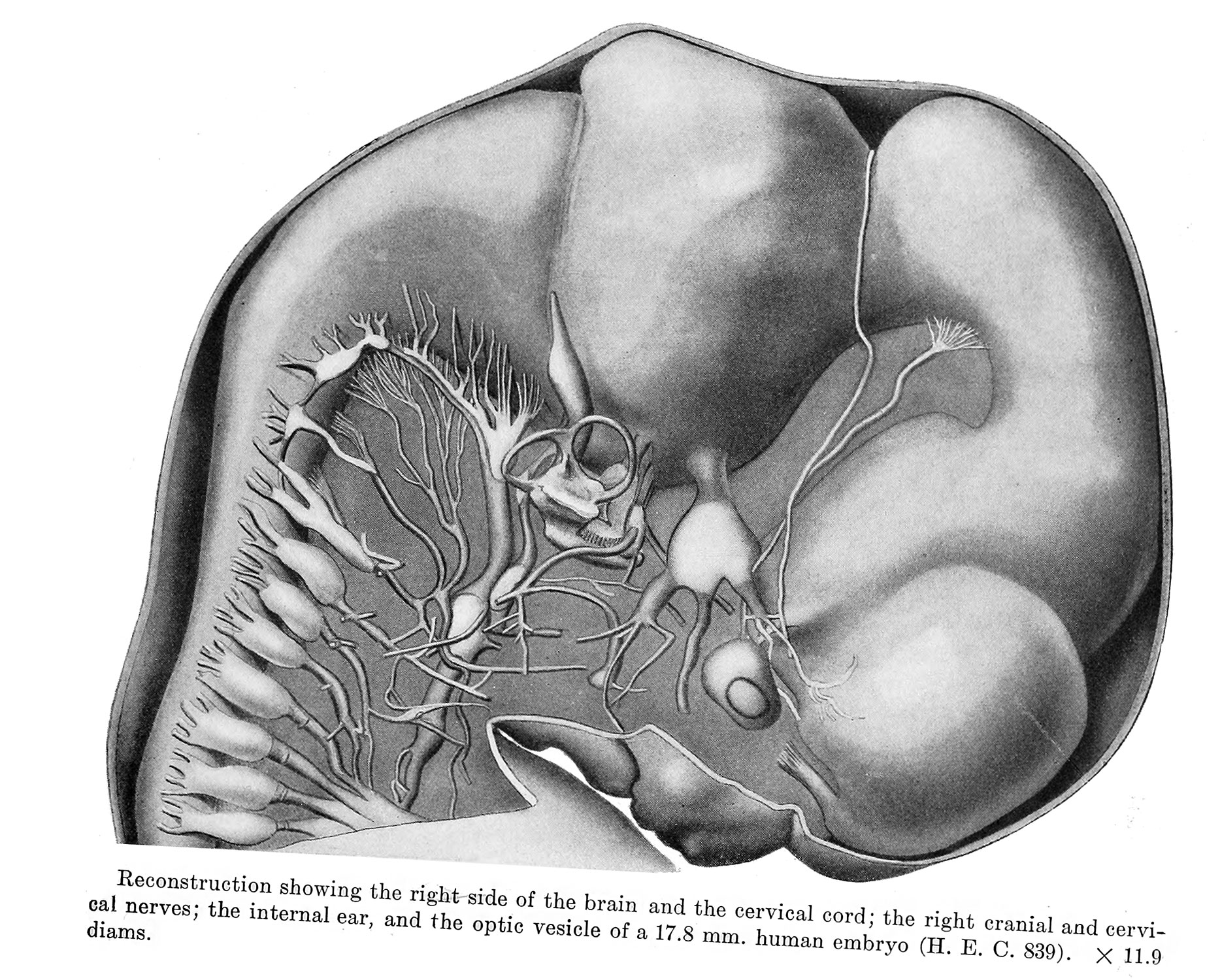 | 1,867 × 1,500 (611 KB) | Z8600021 (talk | contribs) | ==Plate 3== {{Thyng1914 figures}} |
You cannot overwrite this file.
File usage
The following 4 pages use this file: