File:Thyng1914 plate2a.jpg
Original file (1,157 × 1,500 pixels, file size: 338 KB, MIME type: image/jpeg)
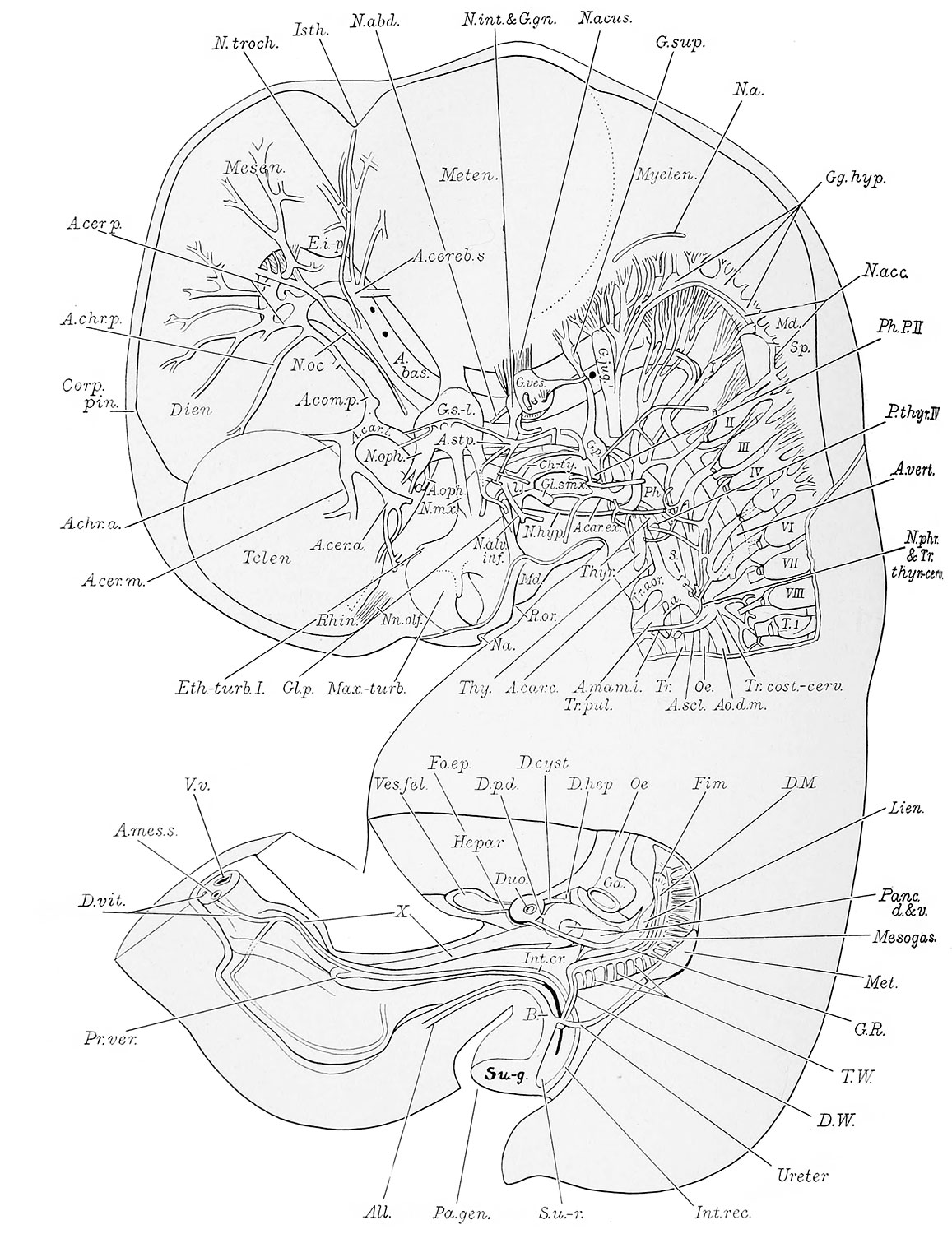
Plate 2
This plate consists of two reconstructions. The upper shows a left lateral view of the brain and cervical cord with the nerves in situ, the aortic arch, and other arteries of the left side of the head and neck. It also represents the oral, nasal and pharyngeal epithelia; the left thymic and thyreoid anlagen; and illustrates in a measure the relation of the nerves and arteries to these epithelial structures. The lower reconstruction shows the pancreas and spleen within the mesogastrium (a portion of the stomach having been removed) ; the left genital ridge, and the left meso- and metanephros with their ducts opening independently into the urogenital division of the cloaca (H. E. C. 839). X 11.2 diams.
- second pharyngeal pouch (Ph.P.2) appears on either side as a low evagination from the lateral pharyngeal wall. It is situated just caudal to the first pouch and projects towards the cephalic aspect of the glossopharyngeal nerve. According to Hammar ('03) this evagination represents only the dorsal part of the primary pouch. A deeply staining cyst is present in this embryo on either side of the pharynx, w^hich evidently belongs to either the ectodermal or entodernial part of the second branchial groove. The left cyst is situated just lateral to the left glossopharyngeal nerve, while the right is just cephalad of the right glossopharyngeal nerve and in close relation to the second pouch. Piersol ('88) found that in rabbit embryos there were formed, in the development of the second pharyngeal pouches, two epithelial tubes on either side, one from the entoderm and the other from the ectodenn, both of which subsequently atrophied. The former, however, persisted longer than the latter. Hammar ('03) described and figured a structure in human embryos protruding above the margin of the tonsilar pouch. In early stages this was connected with the ectoderm, and hence he concluded that it was of ectodermal origin. Fox ('08) did not find any ectodermal remnant in this region of the pig embryo, but described and figured a long filiform process continuous with each of the second pharjmgeal pouches.
- third pharyngeal pouches have lost their connection with the pharynx. They are now represented each by a compact cylinder (Thy., plates 2 and 6) in the side of the neck, and which contains only a slight lumen. The cylinders converge caudally toward the median line and end approxhnately at the level of the aortic arch (Arc.ao.). The right and left cylinders become, eventually, the corresponding lobes of the thymus gland with the exception of the cephalic extremities which are compact epithelial masses (not marked off in the figures) differing in structure from the rest of the anlagen. The cephalic portion of each cylinder is closely applied to the lateral wall of the common carotid artery, and is the part described by Katschenko ('87) as the nodulus thymicus, and by Fox ('08) as the carotid gland. The recent work of Hammar ('11) substantiates the view that this part eventually becomes separated from the thymic cylinder and (coming to lie at the caudo-dorsal border of the lateral part of the thyreoid gland) forms with its fellow of the other side the caudal pair of para-thyreoids. The cephalic ends of the thymic cylinders also show two hollow projections, a medial one, extending toward the pharynx and ending blindly, dorsal to the common carotid artery ; and a more lateral process extending cephalad and ending blindly alongside the ventro-medial surface of the vagus nerve where the latter is crossed on its lateral side by the hypoglossal. The former or medial of these processes is evidently the remains of the thymo-pharyngeal duct. The lateral process seems to be the remains of the cervical sinus fused with the third pharyngeal pouch, as maintained for corresponding structures by Katschenko ('87), Fox ('08), and Hammar ('11), and not an outgrowth from the thymus as conjectured by Prenant ('94) and Bell ('05). Katschenko from a study of this structure in the pig, maintained that it formed a considerable portion of the head of the thymus, a view since corroborated by Prenant ('94) who, however, considers it of entodermal origin. Fox ('08) found that in the cat it apparently atrophied early in development, and that in later stages of the rabbit it had largely, if not entirely disappeared. He is inclined to think that, when it does persist, it does not form an integral part of the thymus, but merely an associated structvire.
- fourth pharyngeal pouch of either side, exclusive of the so-called ultimobranchial (postbranchial) body (Greil '05) is represented by a solid epithelial mass {P-thijr. IV). These masses, which represent the cephalic pair of parathyreoids, are situated dorsal to the lateral lobe of the thyreoid and are entirely separate from the pharynx. That on the right side is bilobate and somewhat removed from the prohferating entoderm of the thyreoid, but on the left the two are intimately connected.
- Embryo 17.8 mm Links: Fig 1 | Fig 2 | Plate 1a | Plate 1b | Plate 2a | Plate 2b | Plate 3a | Plate 3b | Plate 4a | Plate 4b | Plate 5a | Plate 5b | Plate 6 | Harvard Collection | Carnegie stage 19
Reference
Thyng FW. The anatomy of a 17.8 mm human embryo. (1914) Amer. J Anat. 17: 31-112.
Cite this page: Hill, M.A. (2024, April 16) Embryology Thyng1914 plate2a.jpg. Retrieved from https://embryology.med.unsw.edu.au/embryology/index.php/File:Thyng1914_plate2a.jpg
- © Dr Mark Hill 2024, UNSW Embryology ISBN: 978 0 7334 2609 4 - UNSW CRICOS Provider Code No. 00098G
File history
Click on a date/time to view the file as it appeared at that time.
| Date/Time | Thumbnail | Dimensions | User | Comment | |
|---|---|---|---|---|---|
| current | 19:08, 9 April 2014 | | 1,157 × 1,500 (338 KB) | Z8600021 (talk | contribs) |
You cannot overwrite this file.
File usage
The following 7 pages use this file: