File:Thyng1914 plate2a.jpg
Original file (1,157 × 1,500 pixels, file size: 338 KB, MIME type: image/jpeg)
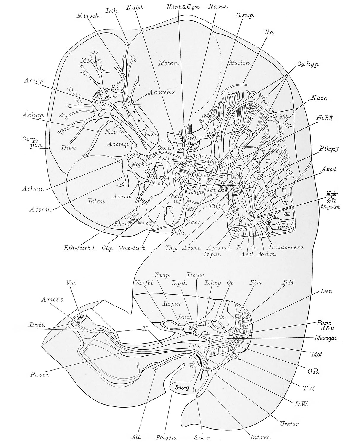
Plate 2
This plate consists of two reconstructions. The upper shows a left lateral view of the brain and cervical cord with the nerves in situ, the aortic arch, and other arteries of the left side of the head and neck. It also represents the oral, nasal and pharyngeal epithelia; the left thymic and thyreoid anlagen; and illustrates in a measure the relation of the nerves and arteries to these epithelial structures. The lower reconstruction shows the pancreas and spleen within the mesogastrium (a portion of the stomach having been removed) ; the left genital ridge, and the left meso- and metanephros with their ducts opening independently into the urogenital division of the cloaca (H. E. C. S39). X 11.2 diams.
- Embryo 17.8 mm Links: Fig 1 | Fig 2 | Plate 1a | Plate 1b | Plate 2a | Plate 2b | Plate 3a | Plate 3b | Plate 4a | Plate 4b | Plate 5a | Plate 5b | Plate 6 | Harvard Collection | Carnegie stage 19
Reference
Thyng FW. The anatomy of a 17.8 mm human embryo. (1914) Amer. J Anat. 17: 31-112.
Cite this page: Hill, M.A. (2024, April 23) Embryology Thyng1914 plate2a.jpg. Retrieved from https://embryology.med.unsw.edu.au/embryology/index.php/File:Thyng1914_plate2a.jpg
- © Dr Mark Hill 2024, UNSW Embryology ISBN: 978 0 7334 2609 4 - UNSW CRICOS Provider Code No. 00098G
File history
Click on a date/time to view the file as it appeared at that time.
| Date/Time | Thumbnail | Dimensions | User | Comment | |
|---|---|---|---|---|---|
| current | 19:08, 9 April 2014 | | 1,157 × 1,500 (338 KB) | Z8600021 (talk | contribs) |
You cannot overwrite this file.
File usage
The following 7 pages use this file: