File:Thyng1914 fig01.jpg: Difference between revisions
(Z8600021 uploaded a new version of "File:Thyng1914 fig01.jpg": no legend) |
mNo edit summary |
||
| Line 1: | Line 1: | ||
==Fig. 1== | ==Fig. 1== | ||
The external features of this embryo are seen in profile view in text figure 1, a reproduction of figure 104 in Minot's (1910) "Laboratory text-book of embryology," also in part in plates 3 and 5. The neck-bend is approximately a right angle; the cephalic flexure is also very nearly a right angled bend, so that the oral aperture is in close proximity to the cardiac region. The dorsal flexure has disappeared almost entirely, only a slight elevation persisting to mark its earlier position. Above this elevation there is a shallow depression, said to disappear in the course of development. | |||
A distinct groove, extending transversely l)etween the medial angles of the developing eyes, separates the forehead from the root of the nose. The maxillary process of either side has joined the adjacent lateral and median nasal processes, obliterating the naso-optic grooves. The nares are open, but separated by a rather low, broad sei)tum. A triangular space still intervenes between the globular processes so that the median region of the upper lip is not well differentiated. The median groove between the ventral ends of the mandibular arches has been obliterated, but differentiation between the chin and lip regions has not occurred. The line of fusion between the first and second branchial arches is marked ventrally by a transverse groove, dorsal to which is seen the fossa conchae. The grooves between the other branchial arches have disappeared. 'A reproduction of figure 104, page 153 of "Laboratory Textbook of Embryology," Charles Sedgwick Minot, edition of 1910, published by P. Blakiston's Son and Company, Pliiladclpliia. The limb buds extend nearly perpendicularly to the longitudinal axis of the body. The upper project slightly beyond the ventral border of the body, and show a differentiation of arm, forearm, and clearly outlined digits. The latter protrude slightly beyond the border of the hand-plate. Upon the lower limb buds are slight indications of developing toes. | |||
Circular thickenings of the epidermis on the lateral body walls mark the developing mammae. In section these thickenings appear slightly convex on the surface, and project into the underlying mesenchyma. The umbilical cord as it leaves the body wall bends towards the right. | |||
Revision as of 10:20, 9 April 2014
Fig. 1
The external features of this embryo are seen in profile view in text figure 1, a reproduction of figure 104 in Minot's (1910) "Laboratory text-book of embryology," also in part in plates 3 and 5. The neck-bend is approximately a right angle; the cephalic flexure is also very nearly a right angled bend, so that the oral aperture is in close proximity to the cardiac region. The dorsal flexure has disappeared almost entirely, only a slight elevation persisting to mark its earlier position. Above this elevation there is a shallow depression, said to disappear in the course of development.
A distinct groove, extending transversely l)etween the medial angles of the developing eyes, separates the forehead from the root of the nose. The maxillary process of either side has joined the adjacent lateral and median nasal processes, obliterating the naso-optic grooves. The nares are open, but separated by a rather low, broad sei)tum. A triangular space still intervenes between the globular processes so that the median region of the upper lip is not well differentiated. The median groove between the ventral ends of the mandibular arches has been obliterated, but differentiation between the chin and lip regions has not occurred. The line of fusion between the first and second branchial arches is marked ventrally by a transverse groove, dorsal to which is seen the fossa conchae. The grooves between the other branchial arches have disappeared. 'A reproduction of figure 104, page 153 of "Laboratory Textbook of Embryology," Charles Sedgwick Minot, edition of 1910, published by P. Blakiston's Son and Company, Pliiladclpliia. The limb buds extend nearly perpendicularly to the longitudinal axis of the body. The upper project slightly beyond the ventral border of the body, and show a differentiation of arm, forearm, and clearly outlined digits. The latter protrude slightly beyond the border of the hand-plate. Upon the lower limb buds are slight indications of developing toes.
Circular thickenings of the epidermis on the lateral body walls mark the developing mammae. In section these thickenings appear slightly convex on the surface, and project into the underlying mesenchyma. The umbilical cord as it leaves the body wall bends towards the right.
- Embryo 17.8 mm Links: Fig 1 | Fig 2 | Plate 1a | Plate 1b | Plate 2a | Plate 2b | Plate 3a | Plate 3b | Plate 4a | Plate 4b | Plate 5a | Plate 5b | Plate 6 | Harvard Collection | Carnegie stage 19
Reference
Thyng FW. The anatomy of a 17.8 mm human embryo. (1914) Amer. J Anat. 17: 31-112.
Cite this page: Hill, M.A. (2024, April 16) Embryology Thyng1914 fig01.jpg. Retrieved from https://embryology.med.unsw.edu.au/embryology/index.php/File:Thyng1914_fig01.jpg
- © Dr Mark Hill 2024, UNSW Embryology ISBN: 978 0 7334 2609 4 - UNSW CRICOS Provider Code No. 00098G
Cite this page: Hill, M.A. (2024, April 16) Embryology Thyng1914 fig01.jpg. Retrieved from https://embryology.med.unsw.edu.au/embryology/index.php/File:Thyng1914_fig01.jpg
- © Dr Mark Hill 2024, UNSW Embryology ISBN: 978 0 7334 2609 4 - UNSW CRICOS Provider Code No. 00098G
File history
Click on a date/time to view the file as it appeared at that time.
| Date/Time | Thumbnail | Dimensions | User | Comment | |
|---|---|---|---|---|---|
| current | 10:08, 9 April 2014 | 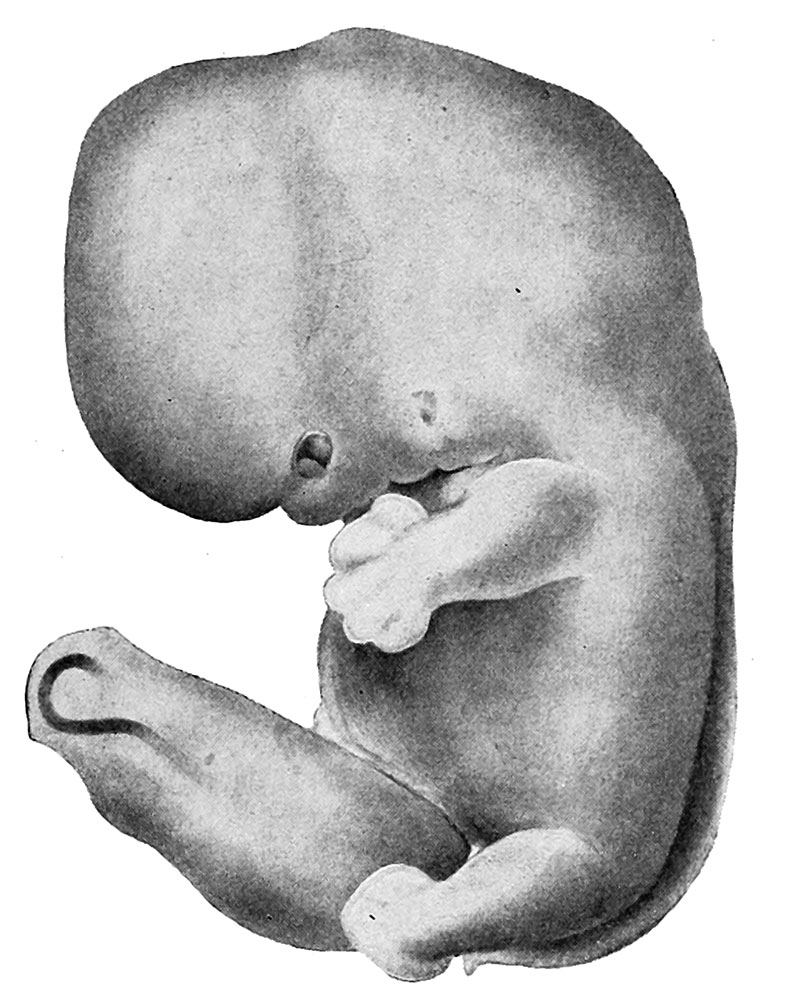 | 786 × 1,000 (140 KB) | Z8600021 (talk | contribs) | no legend |
| 10:04, 9 April 2014 | 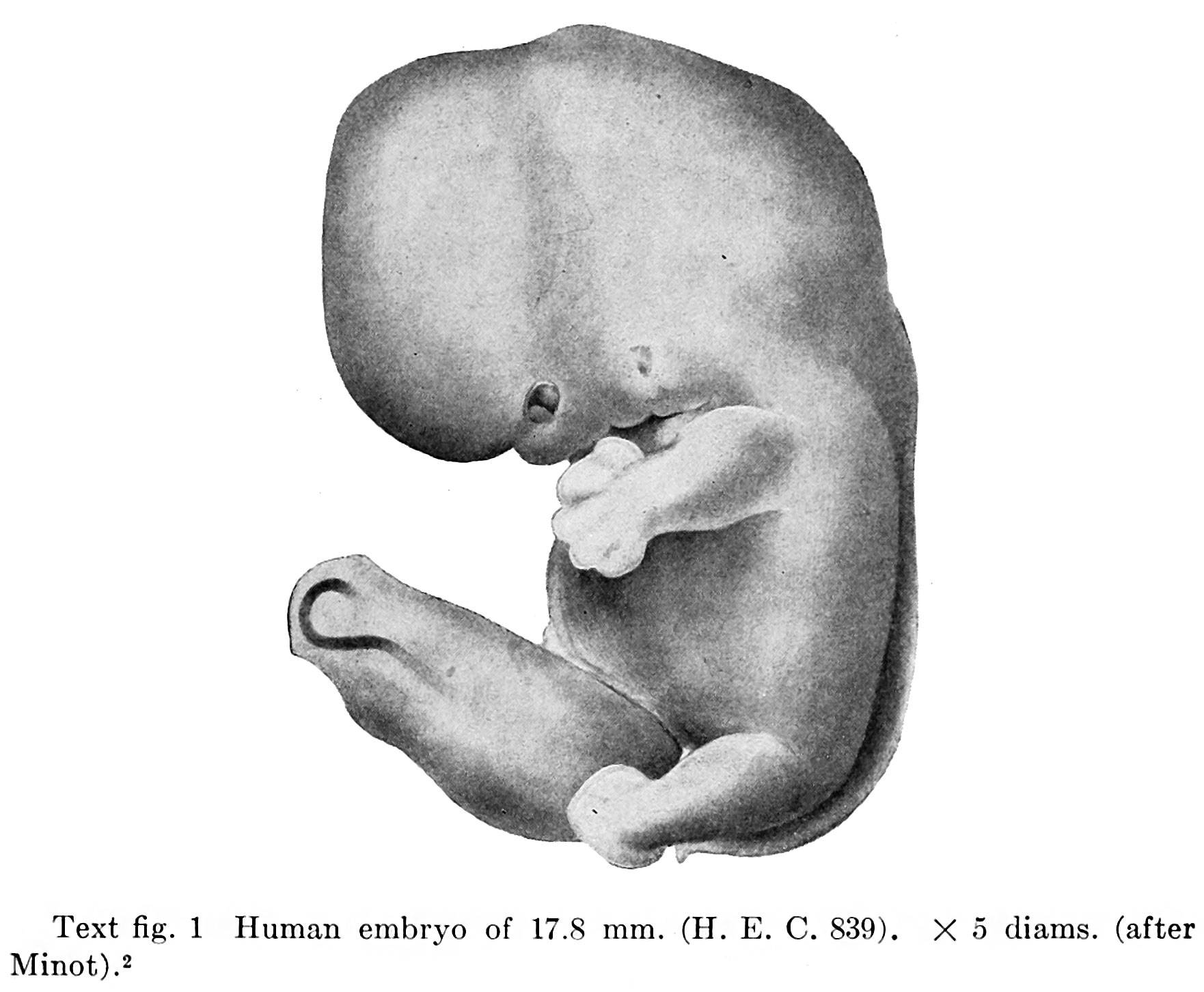 | 1,762 × 1,453 (249 KB) | Z8600021 (talk | contribs) | ==Fig. 1== {{Thyng1914 figures}} |
You cannot overwrite this file.
File usage
The following 4 pages use this file: