File:Tetralogy of fallot-the 4 defects.jpg: Difference between revisions
(This picture demonstrates the four defects (numbers 1-4) found in Tetralogy of Fallot all the 4 defects contribute to poor oxygenation of blood. 1. Pulmonary Stenosis: The small pulmonary vessel causes less blood to enter to the lungs. 2. Displacement o) |
No edit summary |
||
| Line 7: | Line 7: | ||
This is a drawing inspired | This is a drawing inspired and based upon the article "When 'blue babies' grow up: What you need to know about tetralogy of Fallot" by David Fox, Ganesh P Devendra, Stephen A Hart, Richard A Krasuski published in the Cleve Clin J Med in 2010, 77(11);821-8 | ||
PMID:21048055 | PMID:21048055 | ||
Image link: http://www.ccjm.org/content/77/11/821/F1.expansion.html | Image link: http://www.ccjm.org/content/77/11/821/F1.expansion.html | ||
Article link: http://www.ccjm.org/content/77/11/821.long | Article link: http://www.ccjm.org/content/77/11/821.long | ||
Beginning six months after publication, I z3291423 grant the public the non-exclusive right to copy, distribute, or display the Work under a Creative Commons Attribution-Noncommercial-Share Alike 3.0 Unported license, as described at http://creativecommons.org/licenses/by-nc-sa/3.0/ and http://creativecommons.org/licenses/by-nc-sa/3.0/legalcode | |||
--[[User:Z3291423|z3291423]] 12:45, 11 September 2011 (EST) | |||
Revision as of 12:45, 11 September 2011
This picture demonstrates the four defects (numbers 1-4) found in Tetralogy of Fallot all the 4 defects contribute to poor oxygenation of blood.
1. Pulmonary Stenosis: The small pulmonary vessel causes less blood to enter to the lungs. 2. Displacement of the aorta: The displacement allows the aorta to receive both oxygenated and deoxygenated blood. 3. Ventricular Septal Defect: Allows deoxygenated blood to cross from the left side of the heart to the right side, or the opposite way (depending on pressure differences between left and right ventricles). 4. Right Ventricular Hypertrophy: Due to pulmonary stenosis, right ventricle must pump harder to compensare.
This is a drawing inspired and based upon the article "When 'blue babies' grow up: What you need to know about tetralogy of Fallot" by David Fox, Ganesh P Devendra, Stephen A Hart, Richard A Krasuski published in the Cleve Clin J Med in 2010, 77(11);821-8
PMID:21048055 Image link: http://www.ccjm.org/content/77/11/821/F1.expansion.html Article link: http://www.ccjm.org/content/77/11/821.long
Beginning six months after publication, I z3291423 grant the public the non-exclusive right to copy, distribute, or display the Work under a Creative Commons Attribution-Noncommercial-Share Alike 3.0 Unported license, as described at http://creativecommons.org/licenses/by-nc-sa/3.0/ and http://creativecommons.org/licenses/by-nc-sa/3.0/legalcode
--z3291423 12:45, 11 September 2011 (EST)
File history
Click on a date/time to view the file as it appeared at that time.
| Date/Time | Thumbnail | Dimensions | User | Comment | |
|---|---|---|---|---|---|
| current | 12:39, 11 September 2011 | 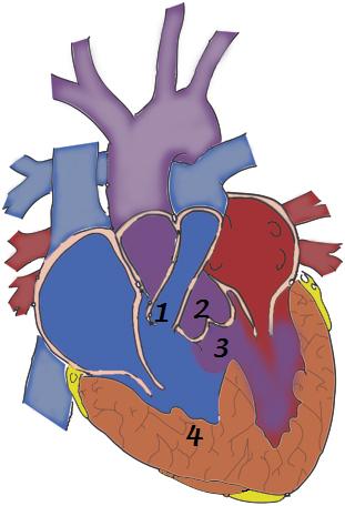 | 311 × 456 (42 KB) | Z3291423 (talk | contribs) | This picture demonstrates the four defects (numbers 1-4) found in Tetralogy of Fallot all the 4 defects contribute to poor oxygenation of blood. 1. Pulmonary Stenosis: The small pulmonary vessel causes less blood to enter to the lungs. 2. Displacement o |
You cannot overwrite this file.
File usage
The following 3 pages use this file: