File:Tetralogy of Fallot 02.jpg
Original file (800 × 796 pixels, file size: 63 KB, MIME type: image/jpeg)
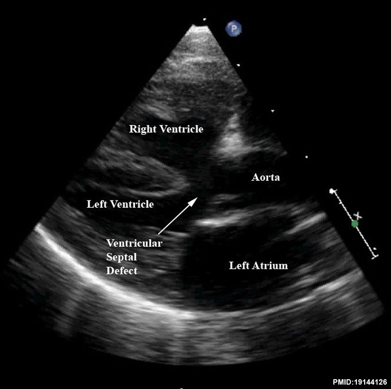
Tetralogy of Fallot Echocardiography
Still frame of a modified parasternal long axis view from patient shows the large ventricular septal defect, aortic override, and right ventricular hypertrophy charactistic of patients with tetralogy of Fallot.
- Links: Tetralogy of Fallot
Reference
<pubmed>19144126</pubmed>| Orphanet J Rare Dis.
Bailliard and Anderson Orphanet Journal of Rare Diseases 2009 4:2 doi:10.1186/1750-1172-4-2
Copyright
© 2009 Bailliard and Anderson; licensee BioMed Central Ltd. This is an Open Access article distributed under the terms of the Creative Commons Attribution License (http://creativecommons.org/licenses/by/2.0), which permits unrestricted use, distribution, and reproduction in any medium, provided the original work is properly cited. Original file name - 1750-1172-4-2-8-l.jpg
File history
Click on a date/time to view the file as it appeared at that time.
| Date/Time | Thumbnail | Dimensions | User | Comment | |
|---|---|---|---|---|---|
| current | 00:05, 23 January 2013 | | 800 × 796 (63 KB) | Z8600021 (talk | contribs) | ==Tetralogy of Fallot Echocardiography == This still frame of a modified parasternal long axis view from the same patient as imaged for Figures 6 and 7 demonstrates the large ventricular septal defect, aortic override, and right ventricular hypertrophy c |
You cannot overwrite this file.
File usage
The following page uses this file: