File:Testis histology 2.jpg
Testis_histology_2.jpg (400 × 500 pixels, file size: 32 KB, MIME type: image/jpeg)
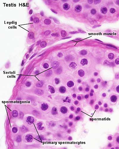
Testis, young and mature - H&E
Identify the capsule and the connective tissue septa extending from it. Identify lobules, convoluted seminiferous tubules and clusters of interstitial cells. The mediastinum testis and rete testis are not visible in all sections.
Original Image Name: tey041he.jpg
Image Source: UWA Blue Histology http://www.lab.anhb.uwa.edu.au/mb140/CorePages/MaleRepro/malerepro.htm#Testes
Links: Histology | Histology Stains | Blue Histology images copyright Lutz Slomianka 1998-2009. The literary and artistic works on the original Blue Histology website may be reproduced, adapted, published and distributed for non-commercial purposes. See also the page Histology Stains.
Cite this page: Hill, M.A. (2024, April 20) Embryology Testis histology 2.jpg. Retrieved from https://embryology.med.unsw.edu.au/embryology/index.php/File:Testis_histology_2.jpg
- © Dr Mark Hill 2024, UNSW Embryology ISBN: 978 0 7334 2609 4 - UNSW CRICOS Provider Code No. 00098G
File history
Click on a date/time to view the file as it appeared at that time.
| Date/Time | Thumbnail | Dimensions | User | Comment | |
|---|---|---|---|---|---|
| current | 16:30, 21 September 2009 | | 400 × 500 (32 KB) | S8600021 (talk | contribs) | Testis, young and mature - H&E Identify the capsule and the connective tissue septa extending from it. Identify lobules, convoluted seminiferous tubules and clusters of interstitial cells. The mediastinum testis and rete testis are not visible in all sec |
You cannot overwrite this file.
File usage
The following 19 pages use this file:
- 2010 BGD Lecture - Development of the Embryo/Fetus 1
- 2010 BGD Practical 3 - Gametogenesis
- 2011 Lab 1 - Gametogenesis
- 2011 Lab 1 - Spermatogenesis
- ANAT2241 Male Reproductive System
- ANAT2341 Lab 1 - Gametogenesis
- ANAT2341 Lab 1 - Spermatogenesis
- BGDA Lecture - Development of the Embryo/Fetus 1
- BGDA Practical - Male Reproductive Tract Histology
- BGDA Practical 3 - Gametogenesis
- BGD Lecture - Sexual Differentiation
- Cell Division - Meiosis
- Lecture - Fertilization
- Lecture - Genital Development
- S
- Sertoli cell
- Spermatozoa Development
- Testis Development
- Talk:BGDA Practical 3 - Gametogenesis