File:TBX1 factor figure.jpg
TBX1_factor_figure.jpg (550 × 351 pixels, file size: 92 KB, MIME type: image/jpeg)
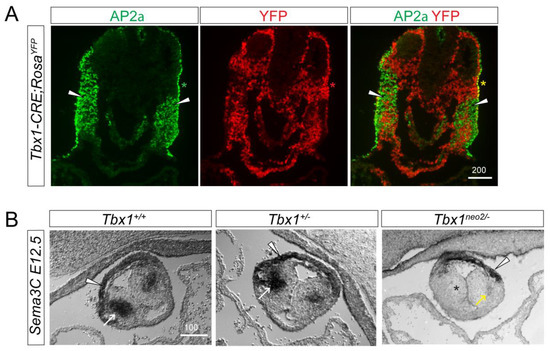
Cardiac neural crest cell defects in Tbx1 mutant embryos. (A) E9.5 Tbx1CRE-RosaYFP embryos were stained for the cNCC marker AP2α together with YFP as a marker for the Tbx1-lineage. cNCCs and Tbx1 do not colocalize (arrowheads). (B) E12.5 Tbx1+/+, Tbx1+/− and Tbx1neo2/− outflow tracts were stained for the cNCC marker Sema3C. cNCCs have migrated into the outflow tract cushions in Tbx1+/+ and Tbx1+/− but not in Tbx1neo2/− embryos. White arrowheads and arrows indicate Sema3C expression in myocardial cuff cells and the septal bridge area, respectively. Asterisk indicates absent Sema3C in the outflow tract right cushion. Yellow arrow marks residual Sema3C-positive cNCCs in the outflow tract left cushion. Scale Bars are in μm.
Reference
Illustration obtained from
Copyright
z5229281
File history
Click on a date/time to view the file as it appeared at that time.
| Date/Time | Thumbnail | Dimensions | User | Comment | |
|---|---|---|---|---|---|
| current | 19:29, 11 October 2018 | | 550 × 351 (92 KB) | Z5229281 (talk | contribs) | Cardiac neural crest cell defects in Tbx1 mutant embryos. (A) E9.5 Tbx1CRE-RosaYFP embryos were stained for the cNCC marker AP2α together with YFP as a marker for the Tbx1-lineage. cNCCs and Tbx1 do not colocalize (arrowheads). (B) E12.5 Tbx1+/+, Tbx1... |
You cannot overwrite this file.
File usage
The following 2 pages use this file: