File:Sudler1902-fig09.jpg: Difference between revisions
From Embryology
mNo edit summary |
(Z8600021 uploaded a new version of File:Sudler1902-fig09.jpg) |
(No difference)
| |
Revision as of 17:49, 21 September 2015
Fig. 9. Lateral view of the model of the nose and pharynx of embryo XLIII
Magnified 15 diameters. I-Iyp., hypophysis; N.,. nasal cavity; Sub. mx. gl., rudiment of submaxillary gland; Thym., thymus; Thyr., thyroid gland; T. p.’, depression caused by the inferior turbinate process; T. p.”, depression ofthe middle turbinate process; V. P.. flrst visceral pouch.
| Historic Disclaimer - information about historic embryology pages |
|---|
 |
- Links: Fig 1 | Fig 2 | Fig 3 | Fig 4 | Fig 5 | Fig 6 | Fig 7 | Fig 8 | Fig 9 | Fig 10 | Fig 11 | Sudler 1902 | Historic Embryology Papers
Reference
Sudler MT. The development of the nose and of the pharynx and its derivatives in man. (1902) Amer. J Anat. 1:391–416.
Cite this page: Hill, M.A. (2024, April 19) Embryology Sudler1902-fig09.jpg. Retrieved from https://embryology.med.unsw.edu.au/embryology/index.php/File:Sudler1902-fig09.jpg
- © Dr Mark Hill 2024, UNSW Embryology ISBN: 978 0 7334 2609 4 - UNSW CRICOS Provider Code No. 00098G
File history
Click on a date/time to view the file as it appeared at that time.
| Date/Time | Thumbnail | Dimensions | User | Comment | |
|---|---|---|---|---|---|
| current | 17:49, 21 September 2015 | 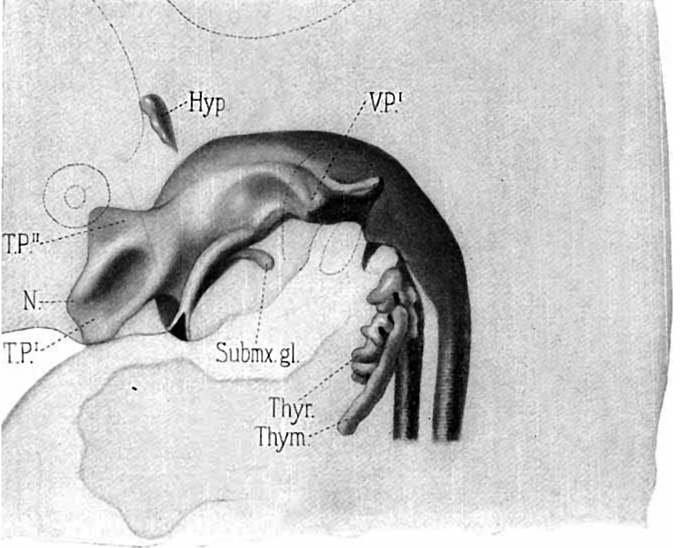 | 1,000 × 798 (124 KB) | Z8600021 (talk | contribs) | |
| 17:49, 21 September 2015 |  | 1,200 × 1,173 (243 KB) | Z8600021 (talk | contribs) | FIG. 9. Lateral view of the model of the nose and pharynx of embryo XLIII. Magnified 15 diameters. I-Iyp., hypophysis; N.,. nasal cavity; Sub. mx. gl., rudiment of submaxillary gland; Thym., thymus; Thyr., thyroid gland; T. p.’, depression caused by... |
You cannot overwrite this file.
File usage
The following 3 pages use this file: