File:Structure of myelinated axons 01.jpg
Original file (651 × 800 pixels, file size: 75 KB, MIME type: image/jpeg)
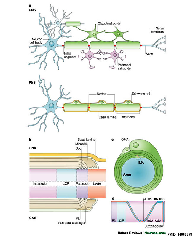
Structure of Myelinated Axons
a - Myelinating glial cells, oligodendrocytes in the central nervous system (CNS) or Schwann cells in the peripheral nervous system (PNS).
- Form the myelin sheath by enwrapping their membrane several times around the axon.
- Myelin covers the axon at intervals (internodes), leaving bare gaps (nodes of Ranvier).
- Oligodendrocytes can myelinate different axons and several internodes per axon.
- Schwann cells myelinate a single internode in a single axon.
b - Schematic longitudinal cut of a myelinated fibre around the node of Ranvier showing a heminode.
- The node, paranode, juxtaparanode (JXP) and internode are labelled.
- The node is contacted by Schwann cell microvilli in the PNS or by processes from perinodal astrocytes in the CNS.
- Myelinated fibres in the PNS are covered by a basal lamina.
- The paranodal loops form a septate-like junction (SpJ) with the axon.
- The juxtaparanodal region resides beneath the compact myelin next to the paranode (PN).
- The internode extends from the juxtaparanodes and lies under the compact myelin.
c - Schematic cross-section of a myelinated nerve depicting the inner and outer mesaxons (IMA and OMA, respectively).
d - Drawing of the specializations found along the internodes.
- A strand composed of paranodal molecules (Caspr, Contactin; red line) flanked by juxtaparanodal proteins (Caspr2, K+ channels and TAG-1; blue lines) extends along the internodal region (the juxtamesaxon) and below the Schmidt–Lanterman incisures (the juxtaincisure).
- In addition, Nf155 and ezrin–radixin–moesin proteins, as well as connexins 29 and 32 are found at the glial side, opposite these axonal strands.
(text from figure legend)
Reference
<pubmed>14682359</pubmed>| Nat Rev Neurosci.
Reprinted by permission from Macmillan Publishers Ltd: Nature Reviews Neuroscience 4, 968-980 (December 2003) | doi:10.1038/nrn1253, copyright (2003)
http://www.nature.com/nrn/journal/v4/n12/fig_tab/nrn1253_F1.html
File history
Click on a date/time to view the file as it appeared at that time.
| Date/Time | Thumbnail | Dimensions | User | Comment | |
|---|---|---|---|---|---|
| current | 09:28, 15 October 2012 | | 651 × 800 (75 KB) | Z8600021 (talk | contribs) | ==Structure of Myelinated Axons== a | Myelinating glial cells, oligodendrocytes in the central nervous system (CNS) or Schwann cells in the peripheral nervous system (PNS), form the myelin sheath by enwrapping their membrane several times around the axon |
You cannot overwrite this file.
File usage
The following 2 pages use this file: