File:Stricht1919b plate2.jpg: Difference between revisions
({{Stricht1919b figures}}) |
mNo edit summary |
||
| Line 1: | Line 1: | ||
== | ==Plate 2== | ||
7 Section tangential to the surface of the organ of Corti, through the third turn of the cochlea. Dog 3 days, 18 hours after birth. Solution of trichloracetic acid 5 per cent in water. Iron hematoxylin, Congo red. | |||
8 Section tangential to the surface of the organ of Corti, through the third turn of the cochlea. Kitten 5 days, 12 hours after birth. Solution of trichloracetic acid 5 per cent in water. Iron hematoxylin, Congo red, light green. | |||
9 Section tangential to the surface of the organ of Corti, through the basal portion of the first (apical) turn of the cochlea. Kitten 12 days after birth. Osn.ic acid 1 per cent aqueous solution for about one hour, followed by immersion in a 5 per cent aqueous solution of trichloracetic acid. Iron hem.atoxylin, Congo red. | |||
10 Vertical spiral (parallel with the spiral rows) section of the organ of Corti, through the second turn of the cochlea. Kitten 11 days after birth. Osmie acid 1 per cent aqueous solution for a^bout one hour, followed by immersion in a 5 per cent aqueous solution of trichloracetic acid. Iron hematoxylin, Congo red. | |||
11 Section tangential to the surface of the organ of Corti, through the basal portion of the second turn of the cochlea. Kitten 12 days after birth. Osmic acid 1 per cent aqueous solution for about half an hour, followed by immersion in a 5 per cent aqueous solution of trichloracetic acid. Iron hematoxylin, Congo red, light green. | |||
12 Section tangential to the surface of the organ of Corti, through the third turn of the cochlea. Kitten 12 days after birth. Osmic acid 1 per cent aqucd»us solution for about half an hour, followed by immersion in a 5 per cent aqueous solution of trichloracetic acid. Iron hematoxylin, Congo red. | |||
{{Stricht1919b figures}} | {{Stricht1919b figures}} | ||
Revision as of 10:37, 19 May 2020
Plate 2
7 Section tangential to the surface of the organ of Corti, through the third turn of the cochlea. Dog 3 days, 18 hours after birth. Solution of trichloracetic acid 5 per cent in water. Iron hematoxylin, Congo red.
8 Section tangential to the surface of the organ of Corti, through the third turn of the cochlea. Kitten 5 days, 12 hours after birth. Solution of trichloracetic acid 5 per cent in water. Iron hematoxylin, Congo red, light green.
9 Section tangential to the surface of the organ of Corti, through the basal portion of the first (apical) turn of the cochlea. Kitten 12 days after birth. Osn.ic acid 1 per cent aqueous solution for about one hour, followed by immersion in a 5 per cent aqueous solution of trichloracetic acid. Iron hem.atoxylin, Congo red.
10 Vertical spiral (parallel with the spiral rows) section of the organ of Corti, through the second turn of the cochlea. Kitten 11 days after birth. Osmie acid 1 per cent aqueous solution for a^bout one hour, followed by immersion in a 5 per cent aqueous solution of trichloracetic acid. Iron hematoxylin, Congo red.
11 Section tangential to the surface of the organ of Corti, through the basal portion of the second turn of the cochlea. Kitten 12 days after birth. Osmic acid 1 per cent aqueous solution for about half an hour, followed by immersion in a 5 per cent aqueous solution of trichloracetic acid. Iron hematoxylin, Congo red, light green.
12 Section tangential to the surface of the organ of Corti, through the third turn of the cochlea. Kitten 12 days after birth. Osmic acid 1 per cent aqucd»us solution for about half an hour, followed by immersion in a 5 per cent aqueous solution of trichloracetic acid. Iron hematoxylin, Congo red.
General Abbreviation
aip, apices of embryonic inner pillar cells
aoh^^, apices or bodies of atrophied hair cells of an outer fourth spiral row
aop, apices or phalanges of the outer pillar cells
cy, cytoplasm in process of cytolysis
c?\ d", d"', apices or bodies of cells of Deiters, respectively, of the first, second, and third rows
H, apex of the cell of Hensen
ih, apices or cell bodies of the inner hair cells
ihd, heads of the inner pillar cells
ij), inner pillars
iph, bodies of the inner pillars
ipl, head-plates of the inner pillars
ipl^, ipV\ respectively, the superficial homogeneous zone and the deeper fibrillated zone of the head plates of the inner pillars
t's', is", apices or cell bodies of supporting cells, respectively, of the first and second inner rows
A, Nerve bundle passing through the foramen nervinum
A'", spiral nerve bundle running between the inner and outer pillar cells or within the tunnel space
iV"', spiral nerve bundle running between the outer pillars and the cells of Deiters of the first row
nd, apices of non-differentiated cells of the greater epithelial ridge
m, nuclei of non-differentiated cells of the greater epithelial ridge
nih, nuclei of inner hair cells
niji, nuclei of inner pillar cells
nis^, nis", nuclei of inner supporting cells, respectively, of the first and second rows
nop, nucleus of an outer pillar cell, seated near the head (abnormality)
oh^, oh", oh}", apices or cell bodies of outer hair cells, respectively, of the first, second, and third rows
ohd, heads of outer pillars
op, outer pillar cells
oph, bodies of outer pillars
oph, phalanx processes of outer pillars
oph^, opd", opd"^, op<^^, respectively, the subphalanx, intercellular, submembraneous segments and the intracephalic roots of the phalanx process of the outer pillar
pd^, pd", pd"^, phalanx processes or apical filaments of cells of Deiters, respectively, of the first, second, and third rows
SN, SN\ the first space of Nuel
t, developing tunnel space
T, tunnel space
tb, terminal bars or obturator septa
VS, vas spirale
- Links: Van der Stricht paper 1919 | Plate 1 | Plate 2 | Plate 3 | organ of Corti
Reference
Van der Stricht O. The development of the pillar cells, tunnel space, and Nuel's spaces in the organ of Corti. (1919) J Comp. Neurol. 30: 283-.
Cite this page: Hill, M.A. (2024, April 19) Embryology Stricht1919b plate2.jpg. Retrieved from https://embryology.med.unsw.edu.au/embryology/index.php/File:Stricht1919b_plate2.jpg
- © Dr Mark Hill 2024, UNSW Embryology ISBN: 978 0 7334 2609 4 - UNSW CRICOS Provider Code No. 00098G
File history
Click on a date/time to view the file as it appeared at that time.
| Date/Time | Thumbnail | Dimensions | User | Comment | |
|---|---|---|---|---|---|
| current | 10:50, 19 May 2020 | 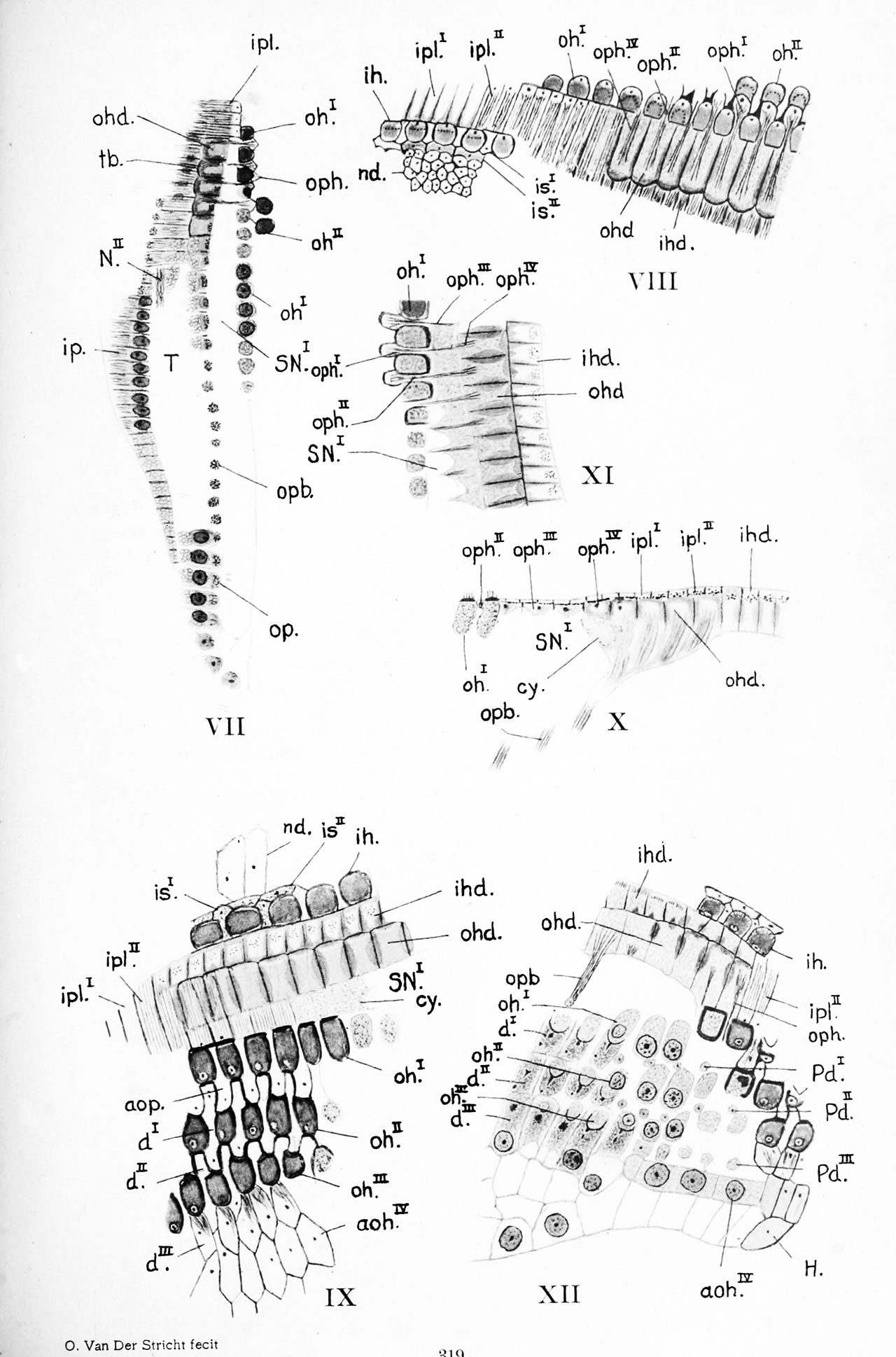 | 1,280 × 1,938 (311 KB) | Z8600021 (talk | contribs) | |
| 10:49, 19 May 2020 |  | 2,255 × 3,414 (759 KB) | Z8600021 (talk | contribs) | ||
| 10:34, 19 May 2020 |  | 2,422 × 3,593 (658 KB) | Z8600021 (talk | contribs) | {{Stricht1919b figures}} |
You cannot overwrite this file.
File usage
The following 2 pages use this file: