File:Streeter1957 fig08.jpg

Original file (1,280 × 1,712 pixels, file size: 507 KB, MIME type: image/jpeg)
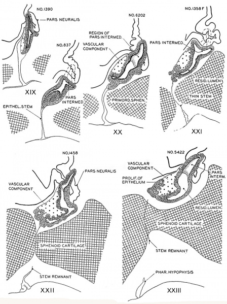
Fig. 8. Drawings of sagittal sections of the hypophysis in embryos of horizons xix to xxiii
Two or more sections were combined in preparing the drawings.
Several sections were utilized in embryos no. 6202, xx, no. 1458, xxii, and no. 5422, xxiii, to represent the lateral processes of the pars intermedia, which grow dorsnlward around the sides of the neural lobe.


| Historic Disclaimer - information about historic embryology pages |
|---|
 |
- Links: 1 Graph Embryos 11-23 | 4 Eye and optic nerve 19-23 | Plate 1 - Cornea | Plate 2 - Hypophysis
Reference
Streeter GL. Developmental Horizons In Human Embryos Description Or Age Groups XIX, XX, XXI, XXII, And XXIII, Being The Fifth Issue Of A Survey Of The Carnegie Collection. (1957) Carnegie Instn. Wash. Publ. 611, Contrib. Embryol., 36: 167-196.
Cite this page: Hill, M.A. (2024, April 23) Embryology Streeter1957 fig08.jpg. Retrieved from https://embryology.med.unsw.edu.au/embryology/index.php/File:Streeter1957_fig08.jpg
- © Dr Mark Hill 2024, UNSW Embryology ISBN: 978 0 7334 2609 4 - UNSW CRICOS Provider Code No. 00098G
File history
Click on a date/time to view the file as it appeared at that time.
| Date/Time | Thumbnail | Dimensions | User | Comment | |
|---|---|---|---|---|---|
| current | 15:03, 8 November 2016 | | 1,280 × 1,712 (507 KB) | Z8600021 (talk | contribs) | |
| 14:58, 8 November 2016 | 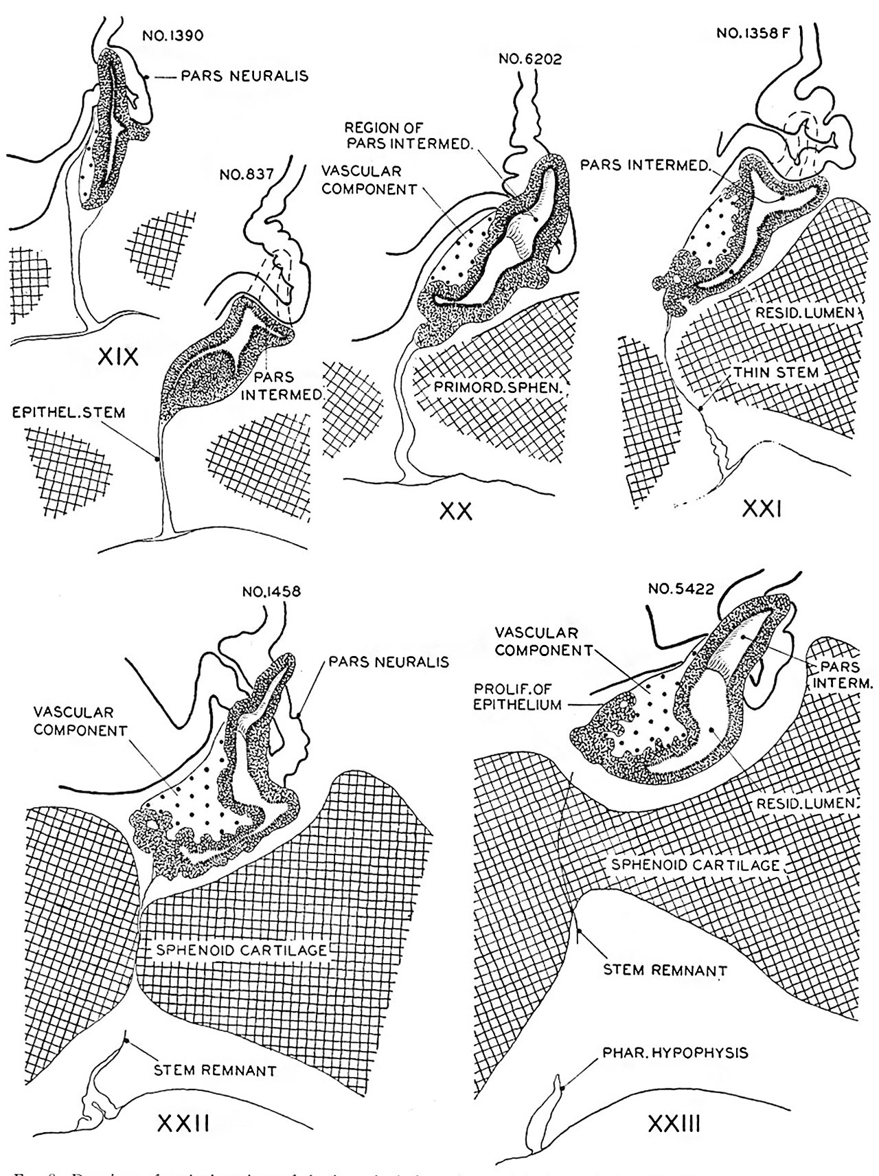 | 1,280 × 1,712 (507 KB) | Z8600021 (talk | contribs) | ||
| 14:57, 8 November 2016 | 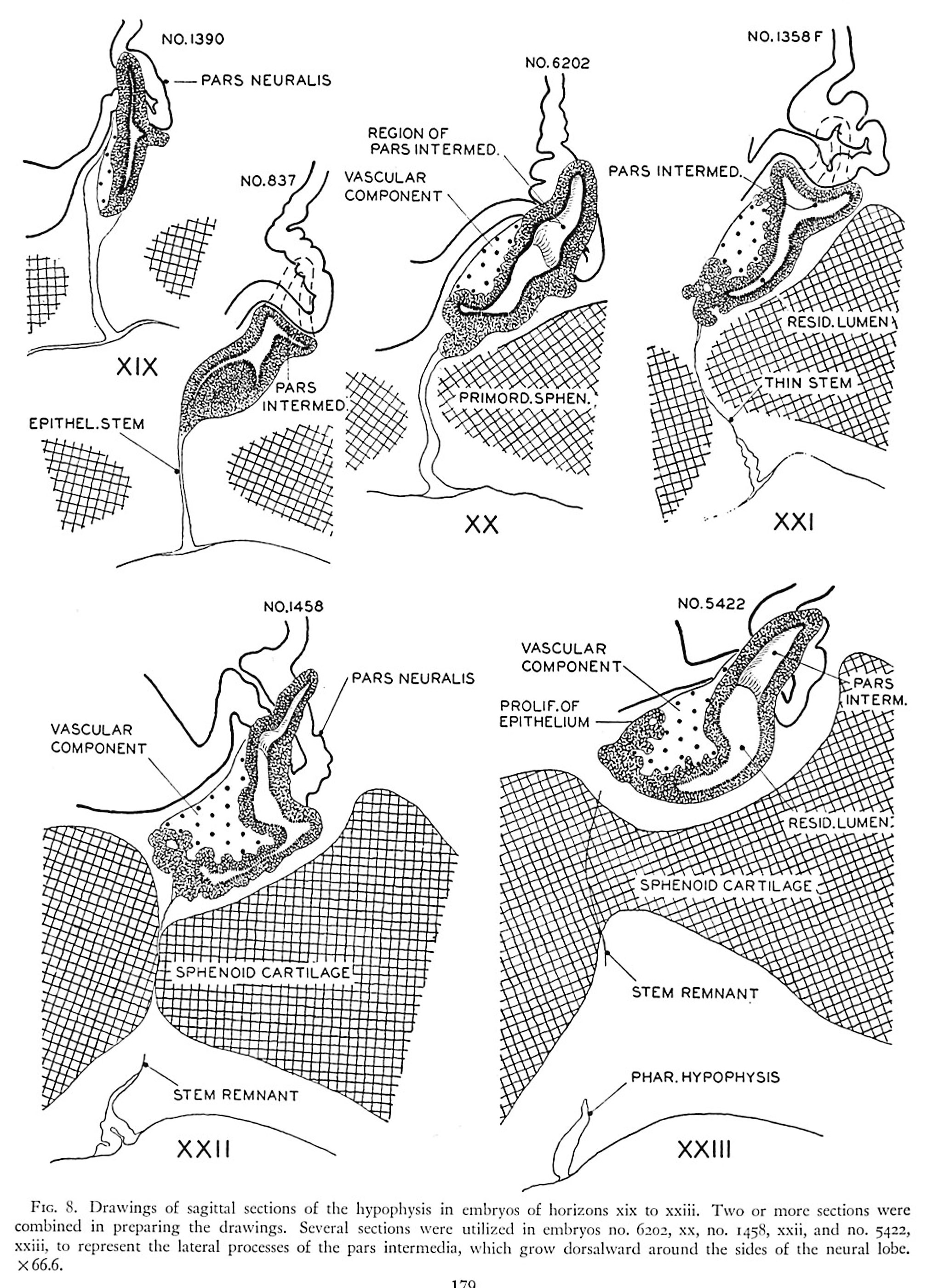 | 2,016 × 2,800 (921 KB) | Z8600021 (talk | contribs) | FIG. 8. Drawings of sngittal sections of the hypophysis in embryos of horizons xix to xxiii. Two or more sections were combined in preparing the drawings. Several sections were utilized in embryos no. 6202, xx, no. 1458, xxii, and no. 342- xxiii, to re... |
You cannot overwrite this file.
File usage
The following 2 pages use this file: