File:Streeter1957 fig06.jpg: Difference between revisions
(==Fig. 6. Drawings of mid-section of the eyeball in embryos of horizons xix to xxiii== The sections were selected to show the organization and relations of parts of the eye in the various age groups. The lens remains nearly spherical throughout this p...) |
mNo edit summary |
||
| (4 intermediate revisions by the same user not shown) | |||
| Line 2: | Line 2: | ||
The sections were selected to show the organization and relations of parts of the eye in the various age groups. The lens remains nearly spherical throughout this period and occupies a relatively large part of the eye. In front of the lens the pupillary membrane develops and the anterior chamber emerges. The hyaloid plexus partly fills the primitive vitreous humor of the eye and forms a network on the posterior surface of the lens. X60. | The sections were selected to show the organization and relations of parts of the eye in the various age groups. The lens remains nearly spherical throughout this period and occupies a relatively large part of the eye. In front of the lens the pupillary membrane develops and the anterior chamber emerges. The hyaloid plexus partly fills the primitive vitreous humor of the eye and forms a network on the posterior surface of the lens. X60. | ||
<gallery> | |||
File:Streeter1957 fig06-19.jpg|[[Carnegie stage 19]] ([[:Category:Carnegie Embryo 409|Embryo 409]]) | |||
File:Streeter1957 fig06-20.jpg|[[Carnegie stage 20]] ([[:Category:Carnegie Embryo 462|Embryo 462]]) | |||
File:Streeter1957 fig06-21.jpg|[[Carnegie stage 21]] ([[:Category:Carnegie Embryo 1358F|Embryo 1358F]]) | |||
File:Streeter1957 fig06-22.jpg|[[Carnegie stage 22]] ([[:Category:Carnegie Embryo 4339|Embryo 4339]]) | |||
File:Streeter1957 fig06-23.jpg|[[Carnegie stage 23]] ([[:Category:Carnegie Embryo 4570|Embryo 4570]]) | |||
</gallery> | |||
<br> | |||
{{Carnegie_stage_table_1}} | |||
<br> | |||
{{Carnegie stage 19 links}} | |||
{{Carnegie stage 20 links}} | |||
{{Carnegie stage 21 links}} | |||
{{Carnegie stage 21 links}} | |||
{{Carnegie stage 23 links}} | |||
<br> | |||
{{Carnegie_stages}} | |||
<br> | |||
{{Streeter1957 figures}} | {{Streeter1957 figures}} | ||
[[Category:Week 8]][[Category:Vision]] | [[Category:Week 8]][[Category:Vision]] | ||
[[Category:Carnegie Embryo 409]][[Category:Carnegie Stage 19]] | |||
[[Category:Carnegie Embryo 462]][[Category:Carnegie Stage 20]] | |||
[[Category:Carnegie Embryo 1358F]][[Category:Carnegie Stage 21]] | |||
[[Category:Carnegie Embryo 4339]][[Category:Carnegie Stage 22]] | |||
[[Category:Carnegie Embryo 4570]][[Category:Carnegie Stage 23]] | |||
Latest revision as of 13:41, 22 May 2017
Fig. 6. Drawings of mid-section of the eyeball in embryos of horizons xix to xxiii
The sections were selected to show the organization and relations of parts of the eye in the various age groups. The lens remains nearly spherical throughout this period and occupies a relatively large part of the eye. In front of the lens the pupillary membrane develops and the anterior chamber emerges. The hyaloid plexus partly fills the primitive vitreous humor of the eye and forms a network on the posterior surface of the lens. X60.


| Week: | 1 | 2 | 3 | 4 | 5 | 6 | 7 | 8 |
| Carnegie stage: | 1 2 3 4 | 5 6 | 7 8 9 | 10 11 12 13 | 14 15 | 16 17 | 18 19 | 20 21 22 23 |
- Carnegie Stages: 1 | 2 | 3 | 4 | 5 | 6 | 7 | 8 | 9 | 10 | 11 | 12 | 13 | 14 | 15 | 16 | 17 | 18 | 19 | 20 | 21 | 22 | 23 | About Stages | Timeline
| Historic Disclaimer - information about historic embryology pages |
|---|
 |
- Links: 1 Graph Embryos 11-23 | 4 Eye and optic nerve 19-23 | Plate 1 - Cornea | Plate 2 - Hypophysis
Reference
Streeter GL. Developmental Horizons In Human Embryos Description Or Age Groups XIX, XX, XXI, XXII, And XXIII, Being The Fifth Issue Of A Survey Of The Carnegie Collection. (1957) Carnegie Instn. Wash. Publ. 611, Contrib. Embryol., 36: 167-196.
Cite this page: Hill, M.A. (2024, April 23) Embryology Streeter1957 fig06.jpg. Retrieved from https://embryology.med.unsw.edu.au/embryology/index.php/File:Streeter1957_fig06.jpg
- © Dr Mark Hill 2024, UNSW Embryology ISBN: 978 0 7334 2609 4 - UNSW CRICOS Provider Code No. 00098G
File history
Click on a date/time to view the file as it appeared at that time.
| Date/Time | Thumbnail | Dimensions | User | Comment | |
|---|---|---|---|---|---|
| current | 14:39, 8 November 2016 | 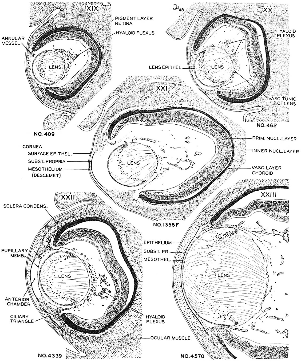 | 1,280 × 1,541 (622 KB) | Z8600021 (talk | contribs) | |
| 14:38, 8 November 2016 | 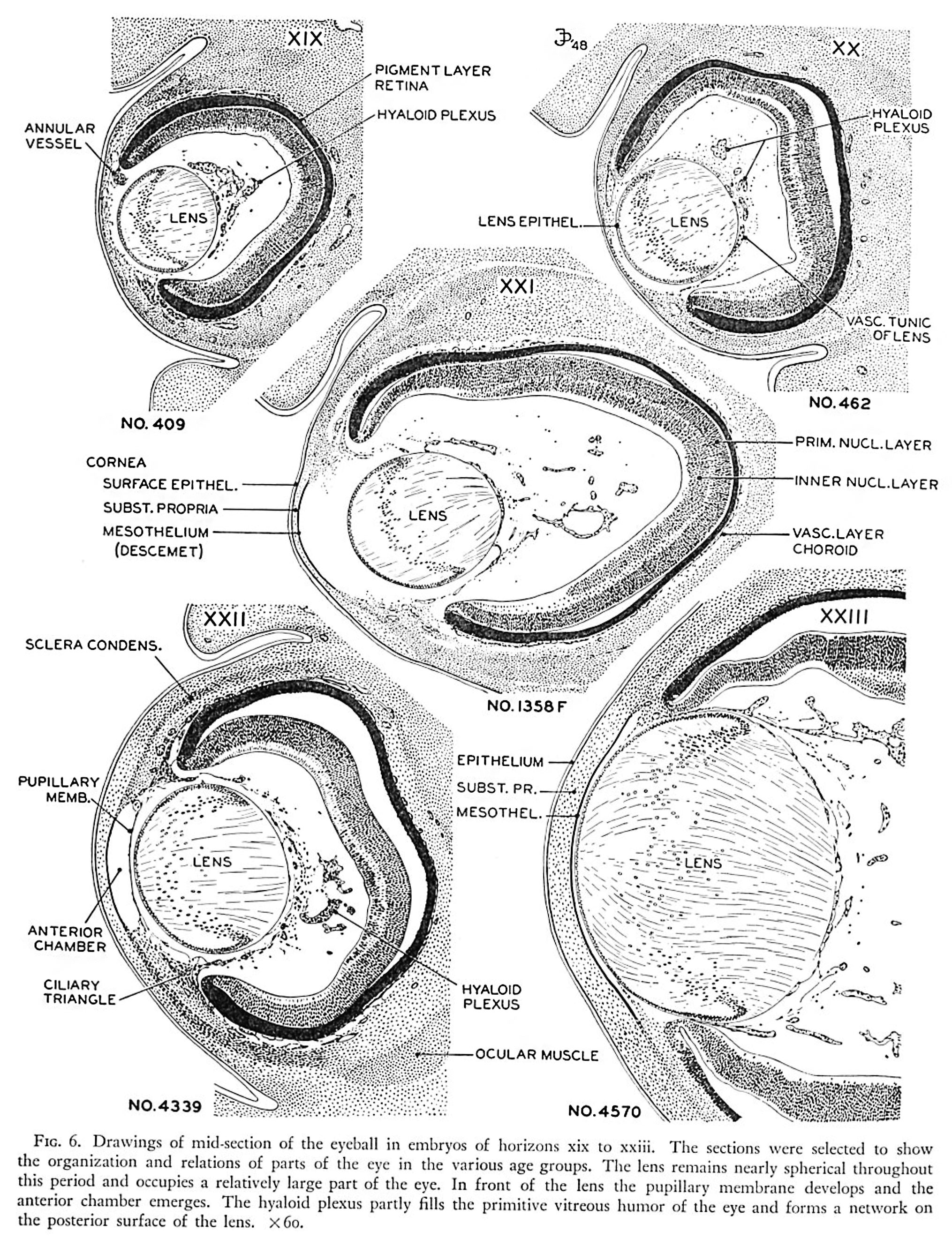 | 2,030 × 2,643 (1.21 MB) | Z8600021 (talk | contribs) | ==Fig. 6. Drawings of mid-section of the eyeball in embryos of horizons xix to xxiii== The sections were selected to show the organization and relations of parts of the eye in the various age groups. The lens remains nearly spherical throughout this p... |
You cannot overwrite this file.
File usage
The following 4 pages use this file: