File:Streeter1922-fig45.jpg

Original file (581 × 781 pixels, file size: 91 KB, MIME type: image/jpeg)
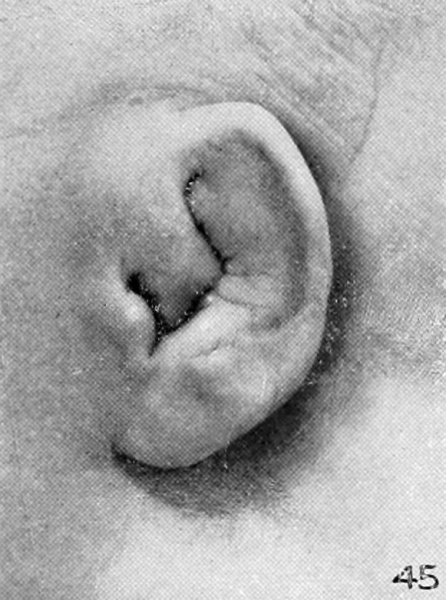
Plate 5. Photographs showing changes occurring in the auricle of the human fetus during the fourth month
The photographs shown on plate 5 represent the changes occurring in the auricle during the fourth month of intrauterine life. As compared with the photographs on plate 4, the principal change is a relative decrease in the size of the crus helicis and tragus. Corresponding to this, it is possible to recognize a conchal cavity which has heretofore been nothing more than a cleft. The concha in the first two photographs (figures 42 and 43) appears to me a little exaggerated, due, probably, to the shrinkage of the auricle. Judging from the preceding and succeeding photographs, the average auricle at this time would be somewhat plumper in appearance. Owing to the fact that these are thinner, one can see for the first time the presence of the plica principalis.
In some cases the right ear was selected and reversed for convenience in comparison. These are indicated by the letter R. All the photographs are taken at an enlargement of 6 diameters.
Specimens are from the Carnegie Collection, and length given is crown-rump.
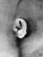
Fig. 42. Embryo No. 1724, 66.2 mm.
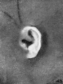
Fig. 43. Embryo No. 2328, 65 mm.
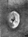
Fig. 44. Embryo No. 2118, 69 mm.
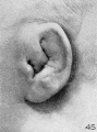
Fig. 45. Embryo No. 981, 85 mm.
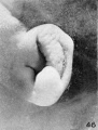
Fig. 46. Embryo No. 1845, 87 mm.
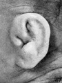
Fig. 47. Embryo No. 1449, 87.3 mm. (R.)
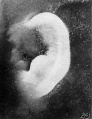
Fig. 48. Embryo No. 2003, 103.5 mm.
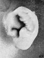
Fig. 49. Embryo No. 1858, 100 mm. (R.)
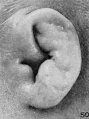
Fig. 50. Embryo No. 2274, 113 mm. (R.)


- In-text Figures: Figure 1 and 2 | Figure 3 and 4 | Figure 5 | Figure 6 and 7 | Figure 8 | Text | Glossary
- Plates: Plate 1 | Plate 2 | Plate 3 | Plate 4 | Plate 5 | Plate 6 | Plates | Glossary
- Figures: 1. Auricle cartilage | 2. External ear | 3. Agnathia | 4. Agnathia+cyclopia | 6. Auricular cartilage embryo 21, 32 and 43 mm | 7. Auricular cartilage 50 mm fetus | 9. Embryo 6 mm | 10. Embryo 12 mm | 11. Embryo 14 mm | 12. Embryo 18 mm | 13. Embryo 1380, 5 mm | 14. Embryo 1767, 11 mm | 15. Embryo 1461, 10 mm | 16. Embryo 562, 13 mm | 17. Embryo 1232, 14 mm | 18. Embryo 475, 15 mm | 19. Embryo 899, 13 mm | 20. Embryo 434, 15 mm | 21. Embryo 492, 16.8 mm | 22. Embryo 576, 17 mm | 23. Embryo 547, 18 mm | 24. Embryo 955, 17 mm | 25. Embryo 1584, 18 mm | 26. Embryo 1134e, 21.3 mm | 27. Embryo 1358b, 33.2 mm | 28. Embryo 1535, 28 mm | 29. Embryo 2163, 36 mm | 30. Embryo 1980, 37 mm | 31. Embryo 1840a, 38.5 mm | 32. Embryo 2075, 40 mm | 33. Embryo 2144, 45.5 mm | 34. Embryo 642, 49 mm | 35. Embryo 2170, 50 mm | 36. Embryo 2095, 52 mm | 37. Embryo 2095, 52 mm | 38. Embryo 2066, 53 mm | 39. Embryo 2079, 56.5 mm. | 40. Embryo 1561, 57 mm | 41. Embryo 218, 62.5 mm. (R.) | 42. Embryo 1724, 66.2 mm | 43. Embryo 2328, 65 mm | 44. Embryo 2118, 69 mm | 45. Embryo 981, 85 mm | 46. Embryo 1845, 87 mm | 47. Embryo 1449, 87.3 mm | 48. Embryo 2003, 103.5 mm | 49. Embryo 1858, 100 mm | 50. Embryo 2274, 113 mm | 51. Embryo 2185, 113.5 mm. | 52. Embryo 9526, 114 mm. | 53. Embryo 1811, 114 mm | 54. Embryo 1716, 119 mm. Fig. 59. 1742, 191.2 mm | 55. Embryo 19576, 119 mm. | 56. Embryo 1782, 135.6 mm | 57. Embryo 1702, 150 mm | 58. Embryo 1708, 154 mm | 59. Embryo 1742, 191.2 mm | Figures
- Related Notes: Outer Ear Development | Carnegie Contributions to Embryology
Reference
Streeter GL. Development of the auricle in the human embryo. (1922) Carnegie Instn. Wash. Publ. 277, Contrib. Embryol., 14: 111-138.
Cite this page: Hill, M.A. (2024, April 18) Embryology Streeter1922-fig45.jpg. Retrieved from https://embryology.med.unsw.edu.au/embryology/index.php/File:Streeter1922-fig45.jpg
- © Dr Mark Hill 2024, UNSW Embryology ISBN: 978 0 7334 2609 4 - UNSW CRICOS Provider Code No. 00098G
| Historic Disclaimer - information about historic embryology pages |
|---|
 |
File history
Click on a date/time to view the file as it appeared at that time.
| Date/Time | Thumbnail | Dimensions | User | Comment | |
|---|---|---|---|---|---|
| current | 08:06, 28 January 2013 | | 581 × 781 (91 KB) | Z8600021 (talk | contribs) | ==Plate 5. Photographs showing changes occurring in the auricle of the human fetus during the fourth month== The photographs shown on plate 5 represent the changes occurring in the auricle during the fourth month of intrauterine life. As compared with th |
You cannot overwrite this file.
File usage
The following 12 pages use this file:
- Book - Contributions to Embryology Carnegie Institution No.69
- File:Streeter1922-fig42.jpg
- File:Streeter1922-fig43.jpg
- File:Streeter1922-fig44.jpg
- File:Streeter1922-fig45.jpg
- File:Streeter1922-fig46.jpg
- File:Streeter1922-fig47.jpg
- File:Streeter1922-fig48.jpg
- File:Streeter1922-fig49.jpg
- File:Streeter1922-fig50.jpg
- File:Streeter1922-plate05.jpg
- Template:Streeter1922 plate5 gallery