File:Streeter1922-fig11.jpg
Original file (667 × 1,000 pixels, file size: 96 KB, MIME type: image/jpeg)
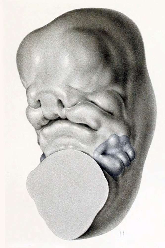
Fig. 11. Reconstruction model of an embryo 14 mm long
No. 940 Carnegie Collection. X 15. Here the parts belonging to the jaw are clearly separated from what are to be the soft parts of the upper neck by a groove, which might be called the mental groove.
Between the stages of 10 and 14 mm. there is rapid progress in the formation of the face, as can be seen by comparing figures 10 and 11.
Figure 11 is drawn from a model to show the details of the face region and the topography of the auricular area, the latter shown in blue. The mouth at this time is fairly well outlined, and one can recognize the region between it and the auricular area which is to form the cheek and jaw. As this region enlarges it will result in the further lateral and dorsal displacement of the auricular area. In the preceding stages the latter still extended downward on the ventral surface of the head, whereas now it is entirely on the lateral surface, and the whole area can be seen in a profile view of the embryo. At this stage the six auricular hillocks show their maximum prominence. The three mandibular hillocks, which at first covered a large part of the mandibular bar, now cover only its caudal margin. The three hyoid hillocks still represent the whole surface of the hyoid bar excepting that part which has been molded into the first cleft. It can be seen in figure 11 that this cleft is much wider than in the younger stages, and we can now speak of a distinct fossa angularis. The ventral third of this fossa becomes relatively deeper to form the external auditory meatus, while the remainder is eventually taken up in the formation of the auricle.
Plate 1. Ventrolateral view of the head in a series of human embryos Fig. 9. Embryo 6 mm | Fig. 10. Embryo 12 mm | Fig. 11. Embryo 14 mm | Fig. 12. Embryo 18 mm
Ventrolateral view of the head in a series of human embryos, showing the change in topography of the auricle in the course of its development. The surface area of the mandibular and hyoid bars entering into the formation of the auricular primordium is colored blue. These figures show the lateral and dorsal migration of the auricle coincident with the formation of the mandible.
- In-text Figures: Figure 1 and 2 | Figure 3 and 4 | Figure 5 | Figure 6 and 7 | Figure 8 | Text | Glossary
- Plates: Plate 1 | Plate 2 | Plate 3 | Plate 4 | Plate 5 | Plate 6 | Plates | Glossary
- Figures: 1. Auricle cartilage | 2. External ear | 3. Agnathia | 4. Agnathia+cyclopia | 6. Auricular cartilage embryo 21, 32 and 43 mm | 7. Auricular cartilage 50 mm fetus | 9. Embryo 6 mm | 10. Embryo 12 mm | 11. Embryo 14 mm | 12. Embryo 18 mm | 13. Embryo 1380, 5 mm | 14. Embryo 1767, 11 mm | 15. Embryo 1461, 10 mm | 16. Embryo 562, 13 mm | 17. Embryo 1232, 14 mm | 18. Embryo 475, 15 mm | 19. Embryo 899, 13 mm | 20. Embryo 434, 15 mm | 21. Embryo 492, 16.8 mm | 22. Embryo 576, 17 mm | 23. Embryo 547, 18 mm | 24. Embryo 955, 17 mm | 25. Embryo 1584, 18 mm | 26. Embryo 1134e, 21.3 mm | 27. Embryo 1358b, 33.2 mm | 28. Embryo 1535, 28 mm | 29. Embryo 2163, 36 mm | 30. Embryo 1980, 37 mm | 31. Embryo 1840a, 38.5 mm | 32. Embryo 2075, 40 mm | 33. Embryo 2144, 45.5 mm | 34. Embryo 642, 49 mm | 35. Embryo 2170, 50 mm | 36. Embryo 2095, 52 mm | 37. Embryo 2095, 52 mm | 38. Embryo 2066, 53 mm | 39. Embryo 2079, 56.5 mm. | 40. Embryo 1561, 57 mm | 41. Embryo 218, 62.5 mm. (R.) | 42. Embryo 1724, 66.2 mm | 43. Embryo 2328, 65 mm | 44. Embryo 2118, 69 mm | 45. Embryo 981, 85 mm | 46. Embryo 1845, 87 mm | 47. Embryo 1449, 87.3 mm | 48. Embryo 2003, 103.5 mm | 49. Embryo 1858, 100 mm | 50. Embryo 2274, 113 mm | 51. Embryo 2185, 113.5 mm. | 52. Embryo 9526, 114 mm. | 53. Embryo 1811, 114 mm | 54. Embryo 1716, 119 mm. Fig. 59. 1742, 191.2 mm | 55. Embryo 19576, 119 mm. | 56. Embryo 1782, 135.6 mm | 57. Embryo 1702, 150 mm | 58. Embryo 1708, 154 mm | 59. Embryo 1742, 191.2 mm | Figures
- Related Notes: Outer Ear Development | Carnegie Contributions to Embryology
Reference
Streeter GL. Development of the auricle in the human embryo. (1922) Carnegie Instn. Wash. Publ. 277, Contrib. Embryol., 14: 111-138.
Cite this page: Hill, M.A. (2024, April 25) Embryology Streeter1922-fig11.jpg. Retrieved from https://embryology.med.unsw.edu.au/embryology/index.php/File:Streeter1922-fig11.jpg
- © Dr Mark Hill 2024, UNSW Embryology ISBN: 978 0 7334 2609 4 - UNSW CRICOS Provider Code No. 00098G
| Historic Disclaimer - information about historic embryology pages |
|---|
 |
File history
Click on a date/time to view the file as it appeared at that time.
| Date/Time | Thumbnail | Dimensions | User | Comment | |
|---|---|---|---|---|---|
| current | 10:44, 27 January 2013 | | 667 × 1,000 (96 KB) | Z8600021 (talk | contribs) | ==Fig. 11. Reconstruction model of an embryo 14 mm long== No. 940 Carnegie Collection. X 15. Here the parts belonging to the jaw are clearly separated from what are to be the soft parts of the upper neck by a groove, which might be called the mental groo |
You cannot overwrite this file.
File usage
The following 5 pages use this file: