File:Streeter1922-01-02.jpg
Original file (1,200 × 668 pixels, file size: 134 KB, MIME type: image/jpeg)
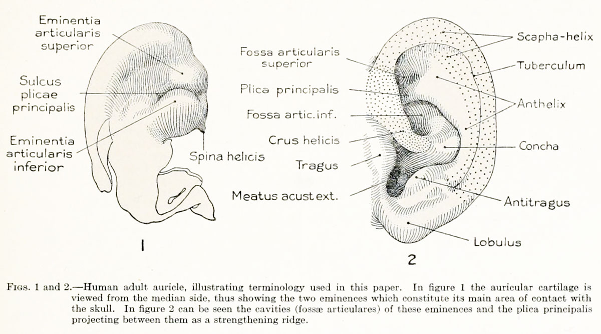
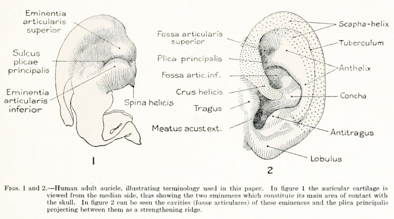
Figs. 1 and 2. Human adult auricle, illustrating terminology used in this paper
In figure 1 the auricular cartilage is viewed from the median side, thus showing the two eminences which constitute its main area of contact with the skull.
In figure 2 can be seen the cavities (fossae articulares) of these eminences and the plica principalis projecting between them as a strengthening ridge.
Terms
- Anthelix - The rounded brim of the concha, from which the secondary part of the. auricle flares out as the scapha-helix. (See fig. 2.)
- Antitragus - The thickened ventral rim of the concha, situated between the incisura intertragica and the anthelix. Apparently a part of the closure mechanism.
- Cavitas conchas - see Concha.
- Concha - The shell-shaped primary part of the auricle immediately surrounding the meatus. As previously used, the term included only the cymba
conchas and the cavitas concha?. In this paper I have extended the term to include also what has been known as the fossa triangularis. The contour of the concha thus is outlined by the tragus, incisura intertragica, antitragus, anthelix, and crus helicis.
- Crus helicis - Formerly restricted to the horizontal portion of the helix, forming a transverse ridge in the floor of the concha. In this paper the term is extended to include all that part of the helix derived from the mandibular arch. (See fig. 2.) It constitutes the lateral free edge of the pars articularis concha?, differing in structure and development from the remainder of the helix.
- Darwin's tubercle - See Tuberculum auricula.
- Eminentia articularis inferior - See Eminentia articularis superior, formerly known as eminentia concha.
- Eminentia articularis superior - Same as eminentia fossae triangularis. The pars articularis of the concha, as viewed from the median side, presents two eminences which constitute the chief area of contact of the auricle with the skull. In this paper these are designated, respectively, eminentia articularis superior and eminentia articularis inferior. (See fig. 1.) The groove between them is the sulcus corresponding to the plica principalis.
- Fossa angularis - [His]. Name applied to the first branchial cleft when modified by the formation of the auricular hillocks, five of which form a plump ring around it.
- Fossa articularis inferior - Same as cymba concha. See Fossa articularis superior.
Fossa articularis superior
Same as fossa triangularis. When the pars articularis concha? is viewed from the lateral side, its floor presents two fossa? (superior and inferior) separated by the plica principalis. (See fig. 2.)
Fossa conchae
[Hammar]. Essentially the same as fossa angularis.
Fossa intercruralis
Same as fossa triangularis, or, as used in this paper, fossa articularis superior.
Fossa scaphoidea
See Scapha.
Fossa triquetra
Same as fossa triangularis, or, as used in this paper, fossa articularis superior.
- Free ear-fold - or freien Ohrfalte [Schwalbe]. The ridge representing first appearance of definitive auricle. Same as helix hyoidalis [Gradenigo], cauda helicis [His], or primitive scapha [Henneberg].
Helix
In adult man the rolled-in margin of the auricle, when viewed as a whole from the lateral side, resembles in outline a coiled spring and on this account it was termed helix. Included under it are parts that are quite different, both embryologically and structurally. Furthermore, it is not applicable to the auricle of other animals. If the term scapha be used for all of the auricle peripheral to the anthelix, and the term helix used for the rolled edge of the scapha, where this occurs, the difficulty is then largely removed. It is so used in this paper, and under scapha-helix will be designated only those parts of the secondary auricle derived from the hyoid arch. The crus helicis is a different structure. The lobulus auricula? is a part of the secondary auricle and bears a similar relation to the concha as does the scapha. (See fig. 2.)
Helix ascendens
[Schwalbe]. The anterior portion of the helix which is derived from the third auricular hillock of the mandibular arch. Partially synonymous with crus helicis, as used by me.
Helix hyoidalis
[Gradenigo]. That portion of the helix derived from the hyoidal arch, from a fold posterior to the fourth, fifth, and sixth auricular hillocks. Same as cauda helicis [His] and helix posterior [Schwalbe].
Helix mandibularis
[Gradenigo]. Fold found in the embryo directly in front of the third auricular hillock and extending down in front of the second and first hillocks. According to Gradenigo, this fold gives origin to the anterior end of the helix, crus helicis, and tragus.
Helix posterior
[Schwalbe]. That portion of the helix derived from the hyoidal arch. Same as helix hyoidalis [Gradenigo].
Incisurae cartilaginis meatus auditorii externi
[Santorini]. Clefts in cartilage of external meatus, somewhat analogous to the clefts between the cartilaginous rings of the trachea.
Incisura intertragica
The cleft between the tragus and antitragus. A derivative of the lower end of the first branchial cleft.
Lamina tragi
Cartilaginous plate supporting tragus.
Lingula auricula
[His]. See Cauda helicis.
Lobulus auriculae
The free edge of the auricle below the antitragus continuous with the scapha helix. See Helix.
Margo oralis helicis
[Baum and Dobers]. Anterior free border of auricle, particularly in such animals as the pig. In a similar way the posterior border is referred to as aboral.
Pars articularis concha
The upper half of the concha. It includes the two articular fossa? (eminentia?), plica principalis, crus helicis, and spina helicis. (See figs. 1 and 2.)
Plica auricularis longitudinalis cranialis
[Henneberg]. The most cranial of the three longitudinal folds of the scapha in such animals as the pig. The others are designated medialis and caudalis, respectively.
Plica principalis
[Boas]. Equivalent to crus inferius anthelicis. Introduced because it is more accurately applied, particularly to the auricle of mammals other than man.
Ponticulus
Ridge on inner surface of conchal cartilage downward from the inferior articular eminence. It appears to be concerned with the ligamentous attachment of the auricle.
Posteron
[Boas]. See Anteron.
Rima helicis
[Albinus]. Perforation of the cartilage of the crus helicis.
Satyr-tip
[Schwalbe]. The tip of the auricle toward the crown of the head. Also called crown-lip or crown-angle.
Scapha
Concave surface of the free portion of the auricle lying between the anthelix and the helix. Term applied by Henneberg to the entire free auricle from the anthelix to the free border. He applies the term helix to the unwrinkled border of the scapha.
Scapha primitiva
[Henneberg]. Same as free ear-fold.
Scutellum
See Scutulum.
Scutulum
(Also known as scutellum, cartilago-scutiformis, or cartilago-sculularis.) This is supposed by some writers to be simply an enlarged spina helicis which has become detached. According to Schmidt, it is an accessory cartilage, connected with the complicated muscular apparatus, which is provided for the auricle of some mammals and is entirely absent in man. See Spina helicis.
Spina helicis
Cartilaginous process extending forward from the pars articularis concha?. (See fig. 1.) It is not in reality a part of the helix. It is supposed that this structure is enlarged and becomes detached in some mammals to form the scutulum.
Taenia lobularis
The fold attaching the lobule to the parotid region. In the embryo it appears before the lobule itself, being derived from the ventral end of the hyoid bar below hillock 6. It is the extension and widening of the taenia as a free fold, to join the lower end of the helix, that produces the lobule.
Torus marginalis
or Randwulst [Henneberg]. The rounded border inclosing the fossa angularis. It makes its appearance as the hillocks disappear. The latter contribute in part to its formation.
Tragus
The thickened margin of the anterior wall of the concha, situated between the incisura intertragica and the crus helicis. Regarded as a part of the closure mechanism.
Tuberculum anthelicis
[His]. Auricular hillock No. 4.
Tuberculum arterius
[His]. Auricular hillock No. 2, the middle hillock of the mandibular arch.
Tuberculum auriculae
The so-called Darwin's tubercle. Corresponds to the true ear-tip of the long-eared mammals [Schwalbe].
Tuberculum centrale
[His]. Transverse elevation in floor of fossa angularis, separating it into an upper and a lower depression, the lower becoming the auditory meatus. It arises as a bulging of the closure plate of the first gill-cleft.
Tuberculum innominatum
Small cartilaginous anterolateral elevation at junction of horizontal portion of crus helicis with the helix ascendens, i. e., mandibular portion of helix.
Tuberculum intermedius
[His]. Auricular hillock No. 3, the one at the top of the first branchial Cleft.
Tuberculum supratragicum
[His]. Term applied to the accessory elevation that sometimes is found at the upper edge of the tragus. In these cases the tragus may be regarded as two-lobed. The separation of the tragus into two lobes occurs in varying degrees of distinctness.
Tuberculum tragicum
[His]. Auricular hillock No. 1, the lowest hillock of the mandibular arch, giving origin to the tragus.
- In-text Figures: Figure 1 and 2 | Figure 3 and 4 | Figure 5 | Figure 6 and 7 | Figure 8 | Text | Glossary
- Plates: Plate 1 | Plate 2 | Plate 3 | Plate 4 | Plate 5 | Plate 6 | Plates | Glossary
- Figures: 1. Auricle cartilage | 2. External ear | 3. Agnathia | 4. Agnathia+cyclopia | 6. Auricular cartilage embryo 21, 32 and 43 mm | 7. Auricular cartilage 50 mm fetus | 9. Embryo 6 mm | 10. Embryo 12 mm | 11. Embryo 14 mm | 12. Embryo 18 mm | 13. Embryo 1380, 5 mm | 14. Embryo 1767, 11 mm | 15. Embryo 1461, 10 mm | 16. Embryo 562, 13 mm | 17. Embryo 1232, 14 mm | 18. Embryo 475, 15 mm | 19. Embryo 899, 13 mm | 20. Embryo 434, 15 mm | 21. Embryo 492, 16.8 mm | 22. Embryo 576, 17 mm | 23. Embryo 547, 18 mm | 24. Embryo 955, 17 mm | 25. Embryo 1584, 18 mm | 26. Embryo 1134e, 21.3 mm | 27. Embryo 1358b, 33.2 mm | 28. Embryo 1535, 28 mm | 29. Embryo 2163, 36 mm | 30. Embryo 1980, 37 mm | 31. Embryo 1840a, 38.5 mm | 32. Embryo 2075, 40 mm | 33. Embryo 2144, 45.5 mm | 34. Embryo 642, 49 mm | 35. Embryo 2170, 50 mm | 36. Embryo 2095, 52 mm | 37. Embryo 2095, 52 mm | 38. Embryo 2066, 53 mm | 39. Embryo 2079, 56.5 mm. | 40. Embryo 1561, 57 mm | 41. Embryo 218, 62.5 mm. (R.) | 42. Embryo 1724, 66.2 mm | 43. Embryo 2328, 65 mm | 44. Embryo 2118, 69 mm | 45. Embryo 981, 85 mm | 46. Embryo 1845, 87 mm | 47. Embryo 1449, 87.3 mm | 48. Embryo 2003, 103.5 mm | 49. Embryo 1858, 100 mm | 50. Embryo 2274, 113 mm | 51. Embryo 2185, 113.5 mm. | 52. Embryo 9526, 114 mm. | 53. Embryo 1811, 114 mm | 54. Embryo 1716, 119 mm. Fig. 59. 1742, 191.2 mm | 55. Embryo 19576, 119 mm. | 56. Embryo 1782, 135.6 mm | 57. Embryo 1702, 150 mm | 58. Embryo 1708, 154 mm | 59. Embryo 1742, 191.2 mm | Figures
- Related Notes: Outer Ear Development | Carnegie Contributions to Embryology
Reference
Streeter GL. Development of the auricle in the human embryo. (1922) Carnegie Instn. Wash. Publ. 277, Contrib. Embryol., 14: 111-138.
Cite this page: Hill, M.A. (2024, April 19) Embryology Streeter1922-01-02.jpg. Retrieved from https://embryology.med.unsw.edu.au/embryology/index.php/File:Streeter1922-01-02.jpg
- © Dr Mark Hill 2024, UNSW Embryology ISBN: 978 0 7334 2609 4 - UNSW CRICOS Provider Code No. 00098G
| Historic Disclaimer - information about historic embryology pages |
|---|
 |
File history
Click on a date/time to view the file as it appeared at that time.
| Date/Time | Thumbnail | Dimensions | User | Comment | |
|---|---|---|---|---|---|
| current | 11:42, 27 January 2013 | | 1,200 × 668 (134 KB) | Z8600021 (talk | contribs) | |
| 23:13, 27 March 2011 | 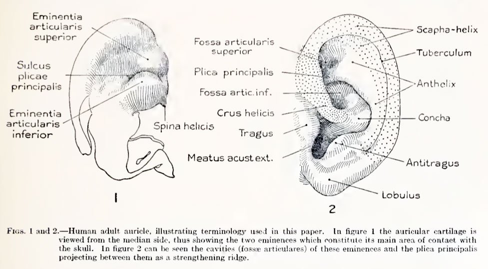 | 977 × 539 (94 KB) | S8600021 (talk | contribs) | {{Streeter1922}} {{Template:Historic Disclaimer}} Category:Human Category:Hearing |
You cannot overwrite this file.
File usage
The following page uses this file: