File:Streeter1921 fig07-09.jpg: Difference between revisions
No edit summary |
No edit summary |
||
| Line 10: | Line 10: | ||
{{Streeter1921}} | {{Streeter1921}} | ||
[[Category:Carnegie Embryo 940]] | |||
[[Category:Carnegie Embryo 349]] | |||
[[Category:Carnegie Embryo 458]] | |||
Revision as of 15:33, 21 April 2012
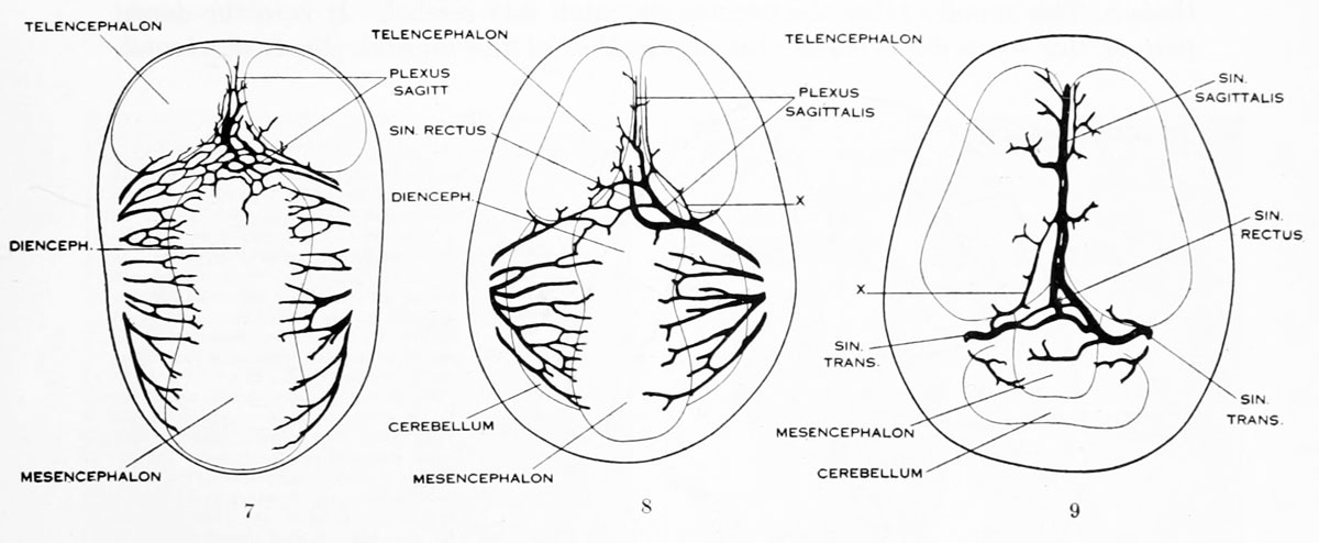
Figures 7, 8. and 9. Three stages in the formation of the sagittal plexus
Three stages in the formation of the sagittal plexus showing its asymmetrical character and its conversion into the superior sagittal sinus. Draining into it from below is the drainage-channel from the chorioidal bodies that is later known as the straight sinus. The channels marked x are interpreted as undergoing retrogression, being replaced by more caudal channels. Large dural channels do not persist in the areas covering the cerebral hemispheres; they are found only in the loose embryonic tissue filling the spaces and fissures that lie between the different subdivsions of the brain.
- Figure 7 is a vertex view of a human embryo 13.8 mm long (Carnegie Collection. No. 940).
- Figure 8 is a vertex view of an embryo 20 mm long (Carnegie Collection, No. 349).
- Figure 9 is a drawing of an injected and cleared specimen 54 mm long (Carnegie Collection, No. 458) reversed.
- 1921 Human Brain Vascular: Fig 1 | Fig 2 | Fig 3 | Fig 4 | Fig 5 | Fig 6 | Fig 7-9 | Fig 10 | Fig 11 | Fig 12 |Fig 13 | Fig 14 | Fig 15 | Fig 16 | Fig 17 | Fig 18 | Fig 19 | Fig 20 | Fig 21 | Fig 22 | Fig 23 | Fig 24 | Fig 25 | Fig 26 | Fig 27 | Plate 1 - embryos 4 mm to birth | Plate 2 - embryo 4 mm | Plate 3 - embryo 11.5 mm | Plate 4 - embryo 21 mm | Plate 5 - embryo 43 mm | Carnegie No.24 | George Streeter
| Historic Disclaimer - information about historic embryology pages |
|---|
 |
Reference
Streeter GL. The developmental alterations in the vascular system of the brain of the human embryo. (1921) Contrib. Embryol., Carnegie Inst. Wash. 8:7-38.
Cite this page: Hill, M.A. (2024, April 25) Embryology Streeter1921 fig07-09.jpg. Retrieved from https://embryology.med.unsw.edu.au/embryology/index.php/File:Streeter1921_fig07-09.jpg
- © Dr Mark Hill 2024, UNSW Embryology ISBN: 978 0 7334 2609 4 - UNSW CRICOS Provider Code No. 00098G
File history
Click on a date/time to view the file as it appeared at that time.
| Date/Time | Thumbnail | Dimensions | User | Comment | |
|---|---|---|---|---|---|
| current | 03:20, 21 April 2012 | 1,200 × 494 (92 KB) | Z8600021 (talk | contribs) | {{Streeter1921}} |
You cannot overwrite this file.
File usage
The following page uses this file: