File:Streeter1917-fig01.jpg: Difference between revisions
mNo edit summary |
mNo edit summary |
||
| (One intermediate revision by the same user not shown) | |||
| Line 3: | Line 3: | ||
(Carnegie Collection, No. 1018). Enlarged 60 diameters. This section shows the topography of the cochlear duct and the general character of the periotic spaces that are developing along its inner margin. Details of this same section as seen under higher magnification are shown in figures 2 and 3. | (Carnegie Collection, No. 1018). Enlarged 60 diameters. This section shows the topography of the cochlear duct and the general character of the periotic spaces that are developing along its inner margin. Details of this same section as seen under higher magnification are shown in figures 2 and 3. | ||
{{Streeter1917}} | {{Streeter1917 figures}} | ||
Latest revision as of 13:21, 16 September 2015
Fig. 1 Section through the second turn of the cochlea in a human fetus 130 mm CR length
(Carnegie Collection, No. 1018). Enlarged 60 diameters. This section shows the topography of the cochlear duct and the general character of the periotic spaces that are developing along its inner margin. Details of this same section as seen under higher magnification are shown in figures 2 and 3.
| Historic Disclaimer - information about historic embryology pages |
|---|
 |
Reference
Streeter GL. The development of the scala tympani, scala vestibuli and perioticular cistern in the human embryo. (1917) Amer. J Anat. 21: 300-320.
Cite this page: Hill, M.A. (2024, April 20) Embryology Streeter1917-fig01.jpg. Retrieved from https://embryology.med.unsw.edu.au/embryology/index.php/File:Streeter1917-fig01.jpg
- © Dr Mark Hill 2024, UNSW Embryology ISBN: 978 0 7334 2609 4 - UNSW CRICOS Provider Code No. 00098G
File history
Click on a date/time to view the file as it appeared at that time.
| Date/Time | Thumbnail | Dimensions | User | Comment | |
|---|---|---|---|---|---|
| current | 08:18, 16 September 2015 | 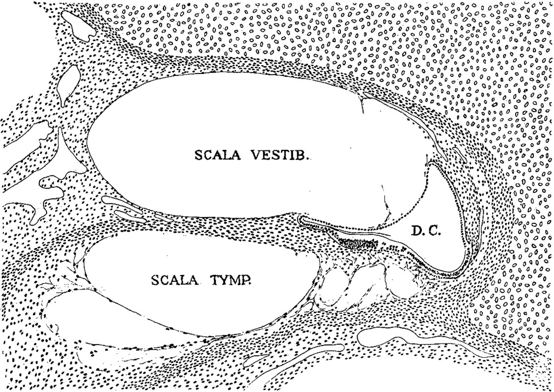 | 1,128 × 800 (298 KB) | Z8600021 (talk | contribs) | |
| 08:16, 16 September 2015 |  | 1,317 × 1,161 (453 KB) | Z8600021 (talk | contribs) | {{Streeter1917}} |
You cannot overwrite this file.
File usage
The following page uses this file: