File:Streeter1908-plate02.jpg
Original file (2,031 × 1,200 pixels, file size: 238 KB, MIME type: image/jpeg)
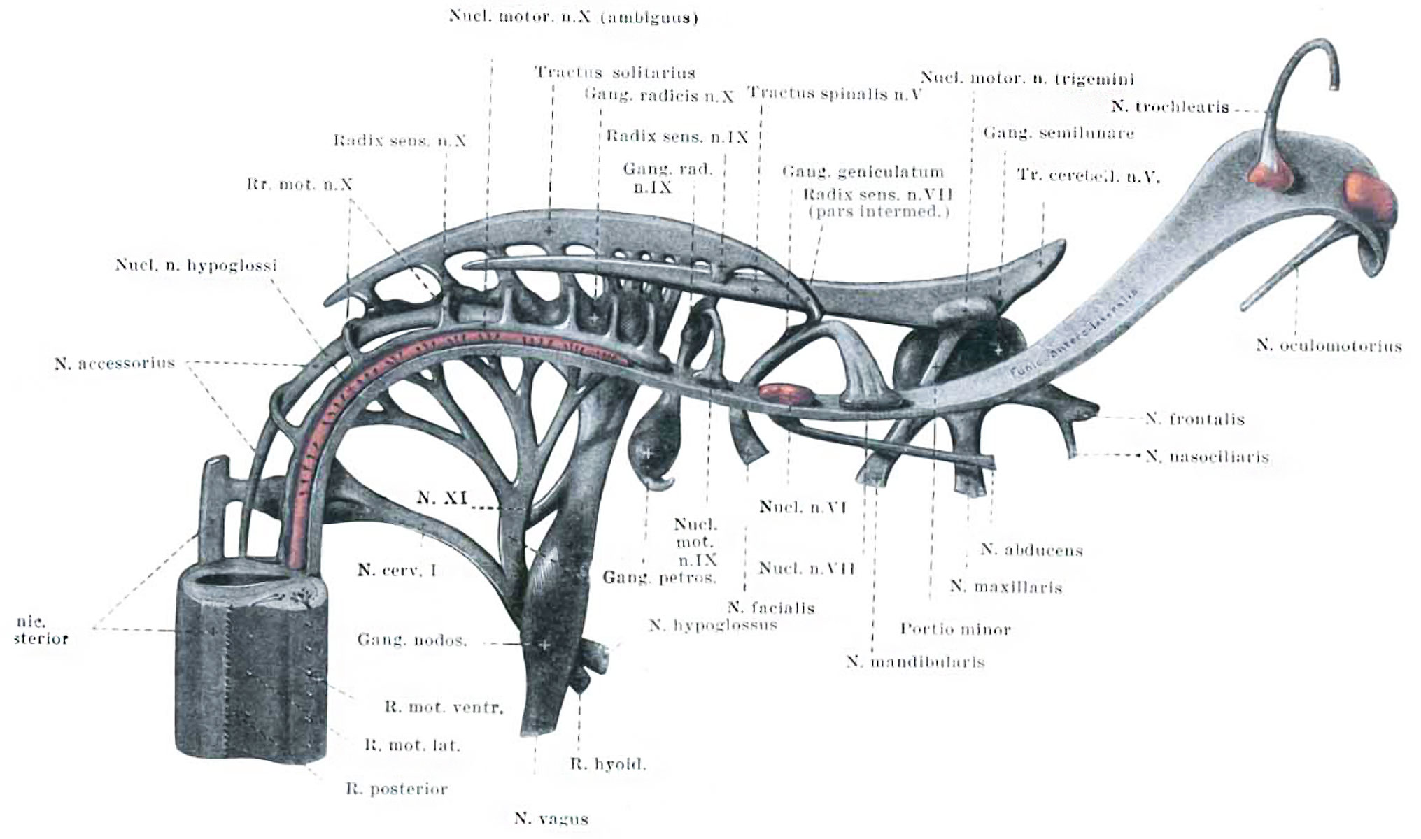
Plate II. Median view of a model of the cranial nerves in the 10 mm Human Embryo
Shown in Plate 1. A portion of the spinal cord is represented and above that everything is cut away, excepting the sensory bundles and motor nuclei of the different nerves, together with that portion of the marginal zone which is to form the funiculus anterolateralis. The somatic motor nuclei are colored red, and it can be seen that they form a column that is practically continuous with the cells of the ventral horn of the spinal cord. Enlarged about 30 diameters.
| Historic Disclaimer - information about historic embryology pages |
|---|
 |
| Online Editor | ||||||||||||||||||
|---|---|---|---|---|---|---|---|---|---|---|---|---|---|---|---|---|---|---|
|
Reference
Streeter GL. The peripheral nervous system in the human embryo at the end of the first month (10 mm) (1908) Amer. J Anat. 8(1): 285–302.
Cite this page: Hill, M.A. (2024, April 19) Embryology Streeter1908-plate02.jpg. Retrieved from https://embryology.med.unsw.edu.au/embryology/index.php/File:Streeter1908-plate02.jpg
- © Dr Mark Hill 2024, UNSW Embryology ISBN: 978 0 7334 2609 4 - UNSW CRICOS Provider Code No. 00098G
File history
Click on a date/time to view the file as it appeared at that time.
| Date/Time | Thumbnail | Dimensions | User | Comment | |
|---|---|---|---|---|---|
| current | 12:16, 14 September 2015 | | 2,031 × 1,200 (238 KB) | Z8600021 (talk | contribs) | |
| 12:09, 14 September 2015 | 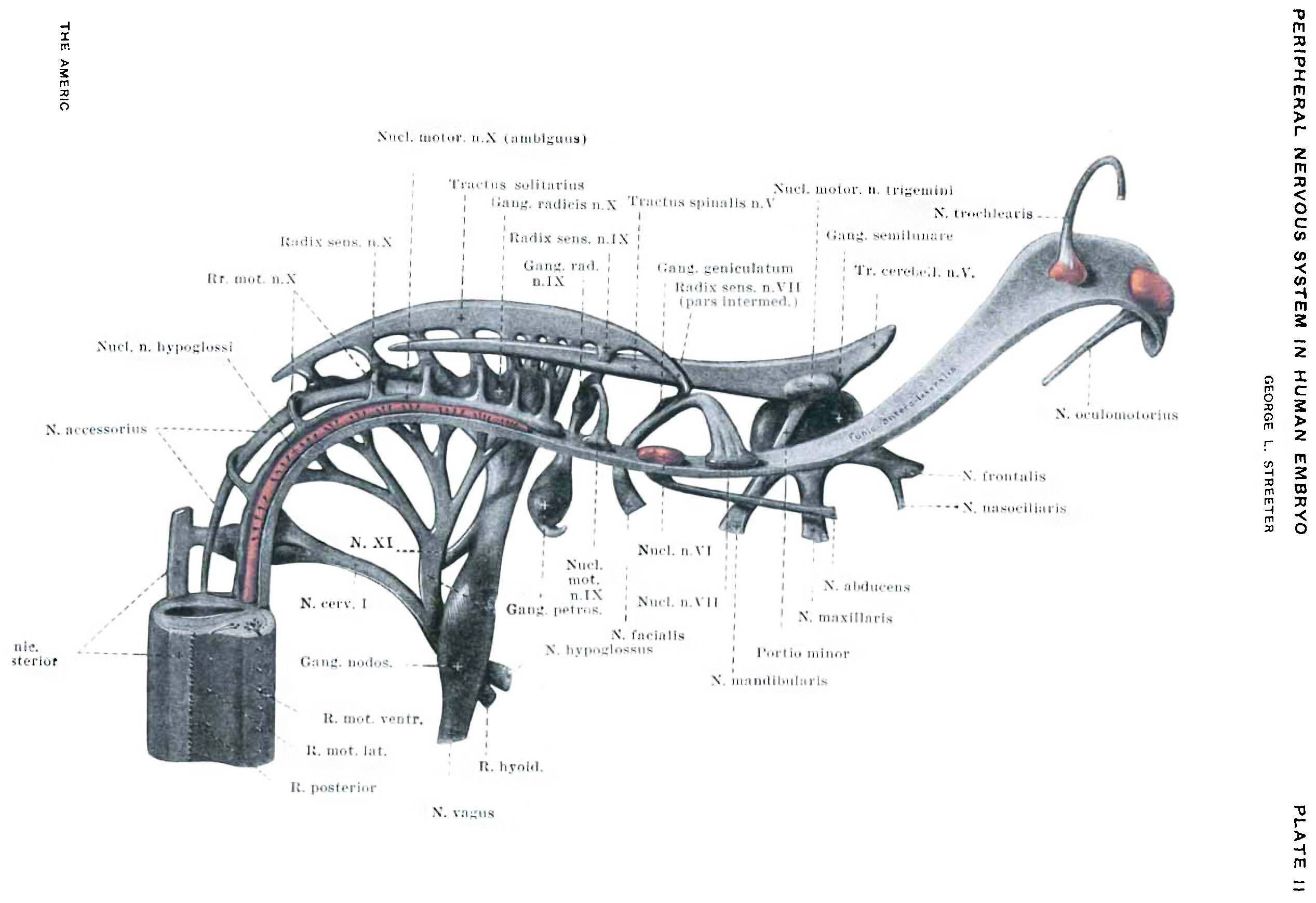 | 2,534 × 1,738 (355 KB) | Z8600021 (talk | contribs) |
You cannot overwrite this file.
File usage
The following 2 pages use this file: