File:Streeter1906 plate01.jpg

Original file (2,708 × 1,786 pixels, file size: 701 KB, MIME type: image/jpeg)
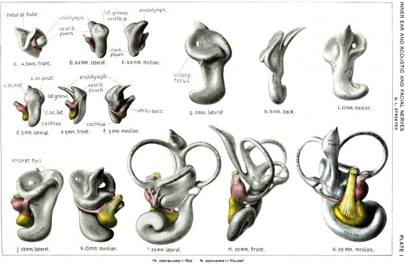
Description of Plates I
The drawings for Plates I and II were prepared under the guidance and assistance of Mr. Max Brodel.
| The reproductions shown. on these plates represent different views, in most cases lateral, front, and median, of seven selected models showing the membranous labyrinth and acoustic complex reconstructed from the following human embryos: No. 148, 4.3 mm.; No. *17, 6.6 mm.; No. 163, 9 mm:; No. 109, 11 mm.; No. 175, 13 mm.; No. 22, 20 mm.; Plate 2 - No. 86, 30 mm.
The colors, yellow and red, are used to indicate respectively the cochlear and vestibular divisions, and in general nerve flbers can be distinguished from ganglion cell masses by their lighter tone.
|
The following abbreviations are used:
|
| Historic Disclaimer - information about historic embryology pages |
|---|
 |
- Mall 1906 Links: Fig 1. 14mm Embryo | Fig 2. 30mm Embryo | Fig 3. Semicircular canal | Fig 4. Membranous Labyrinth | Fig 5. Acoustic nerve complex | Fig 6. Facial-acoustic Complex | Fig 7. Facial Nerve Pig Embryo 20 cm | Fig 8. Geniculate Ganglion | Plate 1. Human Embryo 4 to 20 mm | Plate 2. Human Embryo 30 mm | Membranous Labyrinth and Nerves
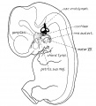
Fig 1 Membranous Labyrinth Human Embryo 14 mm
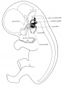
Fig 2 30mm Embryo
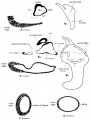
Fig 3 Semicircular canal
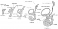
Fig 4 Membranous Labyrinth Growth
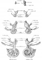
Fig 5 Acoustic nerve complex
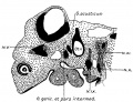
Fig 6 Facial-acoustic Complex Human Embryo 7 mm
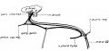
Fig 7 Facial Nerve Pig Embryo 20 cm
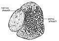
Fig 8 Geniculate Ganglion Human Embryo 30 mm
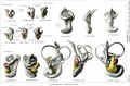
Plate 1. Membranous Labyrinth Human Embryo 4 to 20 mm
Plate 2. Membranous Labyrinth Human Embryo 30 mm


Reference
Streeter GL. On the development of the membranous labyrinth and the acoustic and facial nerves in the human embryo. (1906) Amer. J Anat. 6:139-165.
Cite this page: Hill, M.A. (2024, April 19) Embryology Streeter1906 plate01.jpg. Retrieved from https://embryology.med.unsw.edu.au/embryology/index.php/File:Streeter1906_plate01.jpg
- © Dr Mark Hill 2024, UNSW Embryology ISBN: 978 0 7334 2609 4 - UNSW CRICOS Provider Code No. 00098G
File history
Click on a date/time to view the file as it appeared at that time.
| Date/Time | Thumbnail | Dimensions | User | Comment | |
|---|---|---|---|---|---|
| current | 17:00, 23 July 2015 | | 2,708 × 1,786 (701 KB) | Z8600021 (talk | contribs) | {{Streeter1906 figures}} |
You cannot overwrite this file.
File usage
The following 14 pages use this file:
- BGDB Face and Ear - Late Embryo
- Hearing - Inner Ear Development
- Paper - On the development of the membranous labyrinth and the acoustic and facial nerves in the human embryo
- File:Streeter1906 fig01.jpg
- File:Streeter1906 fig02.jpg
- File:Streeter1906 fig03.jpg
- File:Streeter1906 fig04.jpg
- File:Streeter1906 fig05.jpg
- File:Streeter1906 fig06.jpg
- File:Streeter1906 fig07.jpg
- File:Streeter1906 fig08.jpg
- File:Streeter1906 plate01.jpg
- File:Streeter1906 plate02.jpg
- Template:Streeter1906 figures