File:Streeter-plate04.jpg
From Embryology

Size of this preview: 780 × 600 pixels. Other resolution: 1,301 × 1,000 pixels.
Original file (1,301 × 1,000 pixels, file size: 214 KB, MIME type: image/jpeg)
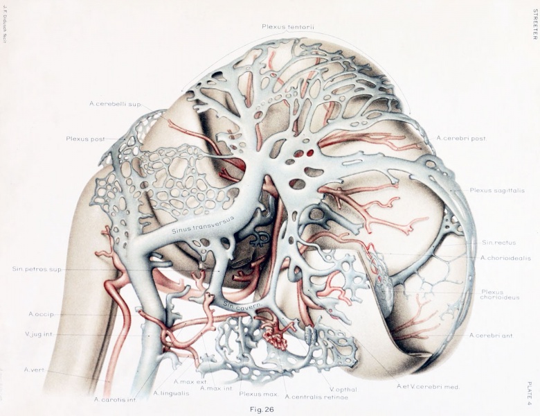
Plate 4. Left lateral view of a wax-plate reconstruction of the larger blood-vessels of the brain in a human embryo 21 mm long
Carnegie Collection Embryo No. 460
Enlarged 16.4 diameters.
- Instead of the head being drained by the primary head-vein, this is now accomplished by a more dorsally situated channel that has formed through the meshes of the middle and posterior dural plexuses to become the transverse sinus.
- Compare with text-figure 3. which shows a left profile of the same specimen.
- All that is left of the primary head-vein is that portion which is to become the cavernous sinus.
- In this model the right cerebral hemisphere has been dissected so as to expose the chorioidal body with its arterial feeder anil the straight sinus draining it.
- The plexiform character of the superior sagittal sinus and of the caudal end of the straight sinus is indicative of their transitory condition.
--Mark Hill 09:37, 17 February 2011 (EST) Estimated as Carnegie Stage 20 Week 8 on basis of CRL. Unknown Shrinkage.
- 1921 Human Brain Vascular: Fig 1 | Fig 2 | Fig 3 | Fig 4 | Fig 5 | Fig 6 | Fig 7-9 | Fig 10 | Fig 11 | Fig 12 |Fig 13 | Fig 14 | Fig 15 | Fig 16 | Fig 17 | Fig 18 | Fig 19 | Fig 20 | Fig 21 | Fig 22 | Fig 23 | Fig 24 | Fig 25 | Fig 26 | Fig 27 | Plate 1 - embryos 4 mm to birth | Plate 2 - embryo 4 mm | Plate 3 - embryo 11.5 mm | Plate 4 - embryo 21 mm | Plate 5 - embryo 43 mm | Carnegie No.24 | George Streeter
| Historic Disclaimer - information about historic embryology pages |
|---|
 |
Reference
Streeter GL. The developmental alterations in the vascular system of the brain of the human embryo. (1921) Contrib. Embryol., Carnegie Inst. Wash. 8:7-38.
Cite this page: Hill, M.A. (2024, April 16) Embryology Streeter-plate04.jpg. Retrieved from https://embryology.med.unsw.edu.au/embryology/index.php/File:Streeter-plate04.jpg
- © Dr Mark Hill 2024, UNSW Embryology ISBN: 978 0 7334 2609 4 - UNSW CRICOS Provider Code No. 00098G
File history
Click on a date/time to view the file as it appeared at that time.
| Date/Time | Thumbnail | Dimensions | User | Comment | |
|---|---|---|---|---|---|
| current | 16:17, 21 April 2012 | | 1,301 × 1,000 (214 KB) | Z8600021 (talk | contribs) | |
| 13:42, 16 February 2011 | 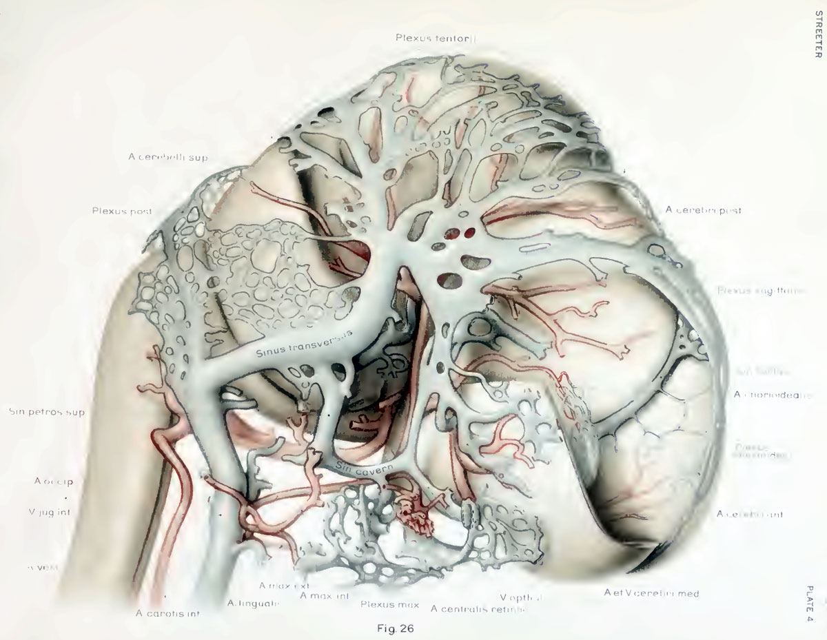 | 1,200 × 929 (160 KB) | S8600021 (talk | contribs) | ==The developmental alterations in the vascular system of the brain of the human embryo== By George L. Streeter. (five plates and twelve text-figures) |
You cannot overwrite this file.