File:Streeter-plate04.jpg: Difference between revisions
No edit summary |
|||
| Line 1: | Line 1: | ||
==Plate 4. | ==Plate 4. The developmental alterations in the vascular system of the brain of the human embryo== | ||
By George L. Streeter. (five plates and twelve text-figures) | By George L. Streeter. (five plates and twelve text-figures) | ||
Left lateral view of a wax-plate reconstruction of the larger blood- | Left lateral view of a wax-plate reconstruction of the larger blood-vessels of the brain in a human embryo 21 mm long (Carnegie Collection, No. 460). Enlarged 16.4 diameters. Instead of the head being drained by the primary head-vein, this is now accomplished by a more dorsally situated channel that has formed through the meshes of the middle and posterior dural plexuses to become the transverse sinus. (Compare with text-figure 3. which shows a left profile of the same specimen.) All that is left of the primary head-vein is that portion which is to become the cavernous sinus. In this model the right cerebral hemisphere has been dissected so as to expose the chorioidal body with its arterial feeder anil the straight sinus draining it. The plexiform character of the superior sagittal sinus and of the caudal end | ||
of the straight sinus is indicative of their transitory condition. | of the straight sinus is indicative of their transitory condition. | ||
--[[User:S8600021|Mark Hill]] 09:37, 17 February 2011 (EST) Estimated as Carnegie Stage 20 Week 8 on basis of CRL. Unknown Shrinkage. | |||
[[Category:Human]] [[Category:Head]] [[Category:Cardiovascular]] | [[Category:Human]] [[Category:Head]] [[Category:Cardiovascular]] [[Category:Carnegie Stage 20]] [[Category:Week 8]] | ||
Revision as of 08:37, 17 February 2011
Plate 4. The developmental alterations in the vascular system of the brain of the human embryo
By George L. Streeter. (five plates and twelve text-figures)
Left lateral view of a wax-plate reconstruction of the larger blood-vessels of the brain in a human embryo 21 mm long (Carnegie Collection, No. 460). Enlarged 16.4 diameters. Instead of the head being drained by the primary head-vein, this is now accomplished by a more dorsally situated channel that has formed through the meshes of the middle and posterior dural plexuses to become the transverse sinus. (Compare with text-figure 3. which shows a left profile of the same specimen.) All that is left of the primary head-vein is that portion which is to become the cavernous sinus. In this model the right cerebral hemisphere has been dissected so as to expose the chorioidal body with its arterial feeder anil the straight sinus draining it. The plexiform character of the superior sagittal sinus and of the caudal end
of the straight sinus is indicative of their transitory condition.
--Mark Hill 09:37, 17 February 2011 (EST) Estimated as Carnegie Stage 20 Week 8 on basis of CRL. Unknown Shrinkage.
File history
Click on a date/time to view the file as it appeared at that time.
| Date/Time | Thumbnail | Dimensions | User | Comment | |
|---|---|---|---|---|---|
| current | 16:17, 21 April 2012 | 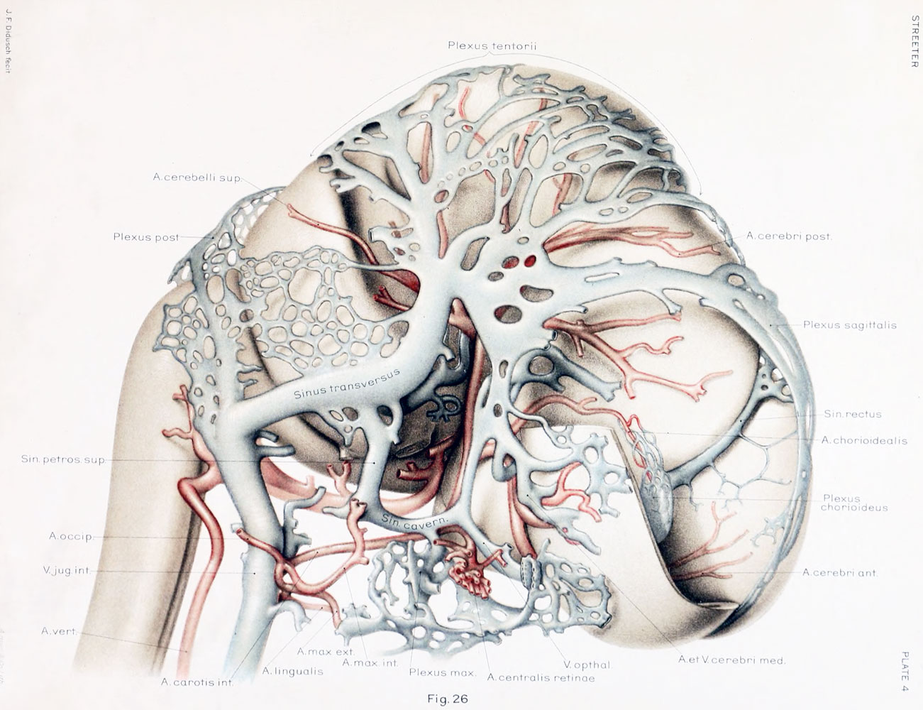 | 1,301 × 1,000 (214 KB) | Z8600021 (talk | contribs) | |
| 13:42, 16 February 2011 | 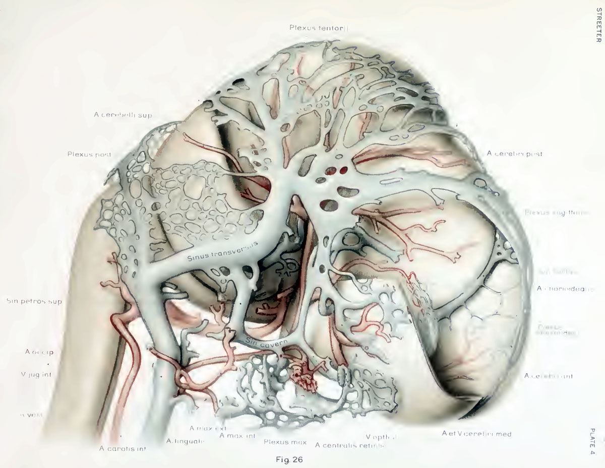 | 1,200 × 929 (160 KB) | S8600021 (talk | contribs) | ==The developmental alterations in the vascular system of the brain of the human embryo== By George L. Streeter. (five plates and twelve text-figures) |
You cannot overwrite this file.