File:Streeter-plate03.jpg

Original file (1,398 × 1,000 pixels, file size: 192 KB, MIME type: image/jpeg)
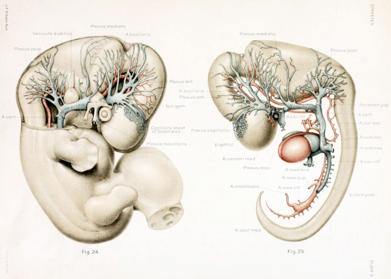
Plate 3. The developmental alterations in the vascular system of the brain of the human embryo
By George L. Streeter. (five plates and twelve text-figures)
ight and left profile views of a wax-plate reconstruction of the blood-vessels of the brain in a liuman embryo 11.5 mm. long (Carnegie Collection. No. 544). Enlarged about 14 diameters. The primary head-vein still constitutes the main drainage-channel of the head. The manner in which its tributaries tap the deep capillary sheet investing the brain is indicated over a small area of the cerebral hemisphere. A capillary mesh of that kind invests the entire central nervous system, but is not shown in the model.
--Mark Hill 09:42, 17 February 2011 (EST) Estimated as Carnegie Stage 17 Week 6 on the basis of CRL and the presence of digital rays.
File history
Click on a date/time to view the file as it appeared at that time.
| Date/Time | Thumbnail | Dimensions | User | Comment | |
|---|---|---|---|---|---|
| current | 09:44, 21 April 2012 | | 1,398 × 1,000 (192 KB) | Z8600021 (talk | contribs) | |
| 13:42, 16 February 2011 |  | 1,200 × 862 (119 KB) | S8600021 (talk | contribs) | ==The developmental alterations in the vascular system of the brain of the human embryo== By George L. Streeter. (five plates and twelve text-figures) |
You cannot overwrite this file.
File usage
The following 3 pages use this file: