File:Streeter-plate01.jpg
From Embryology
Size of this preview: 757 × 599 pixels. Other resolution: 1,200 × 950 pixels.
Original file (1,200 × 950 pixels, file size: 138 KB, MIME type: image/jpeg)
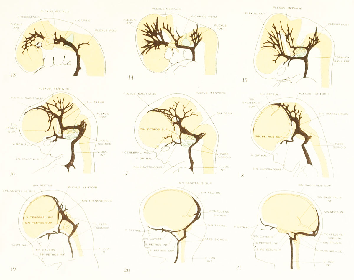
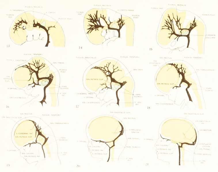
Plate 1. The developmental alterations in the vascular system of the brain of the human embryo
By George L. Streeter. (five plates and twelve text-figures)
Simplified profile drawings of the dural veins, showing the manner in which they adapt themselves to the growth and change
in form of the brain in human embryos from 4 mm. to birth.
- Fig. 13, embryo No. 588, 4 mm
- Fig. 14, embryo No. 940, 14 mm
- Fig. 15, embryo No. 144. 18 mm
- Fig. 16, embryo No. 460, 21 mm
- Fig. 17, embryo No. 632, 24 mm
- Fig. 18, embryo No. 199, 35 mm
- Fig. 19, embryo No. 96, 50 mm CR length
- Fig. 20, embryo No. 234a, 80 mm CR length
- Fig. 21, adult
File history
Click on a date/time to view the file as it appeared at that time.
| Date/Time | Thumbnail | Dimensions | User | Comment | |
|---|---|---|---|---|---|
| current | 04:03, 21 April 2012 | | 1,200 × 950 (138 KB) | Z8600021 (talk | contribs) | |
| 13:41, 16 February 2011 |  | 1,149 × 912 (153 KB) | S8600021 (talk | contribs) | ==The developmental alterations in the vascular system of the brain of the human embryo== By George L. Streeter. (five plates and twelve text-figures) |
You cannot overwrite this file.
File usage
The following 2 pages use this file: