File:Strayer1943 fig18.jpg: Difference between revisions
From Embryology
(Fig. 18. Photomicrograph low power C.H. Embryo 10. length 167 mm., slide 245. Section with the femur in 90° flexion and external rotation. The head of the femur has been thrown inferiorly and laterally so that its greatest diameter is out of the circl...) |
(Z8600021 uploaded a new version of File:Strayer1943 fig18.jpg) |
(No difference)
| |
Latest revision as of 14:18, 9 November 2019
Summary
Fig. 18. Photomicrograph low power C.H. Embryo 10. length 167 mm., slide 245. Section with the femur in 90° flexion and external rotation. The head of the femur has been thrown inferiorly and laterally so that its greatest diameter is out of the circle of the glenoid labrum and cotyloid ligament. The inferior capsule is stretched and bulging. If force were applied in the long axis of the femur, the head would be forced outside the glenoid labrum to cause inferior dislocation.
Reference
Strayer MMJr. The embryology of the human hip joint. (1943) Yale J Biol. Med. 16(1): 13–26.6. PMCID: PMC2601352
File history
Click on a date/time to view the file as it appeared at that time.
| Date/Time | Thumbnail | Dimensions | User | Comment | |
|---|---|---|---|---|---|
| current | 14:18, 9 November 2019 | 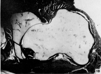 | 325 × 238 (28 KB) | Z8600021 (talk | contribs) | |
| 14:17, 9 November 2019 |  | 369 × 581 (76 KB) | Z8600021 (talk | contribs) | Fig. 18. Photomicrograph low power C.H. Embryo 10. length 167 mm., slide 245. Section with the femur in 90° flexion and external rotation. The head of the femur has been thrown inferiorly and laterally so that its greatest diameter is out of the circl... |
You cannot overwrite this file.
File usage
The following page uses this file: